Clinical Areas:

Retina and Fundus

Glaucoma and Intraocular Pressure

Myopia Control

Cataract

  • Home
  • Miis Fundus Camera

MiiS Fundus Camera

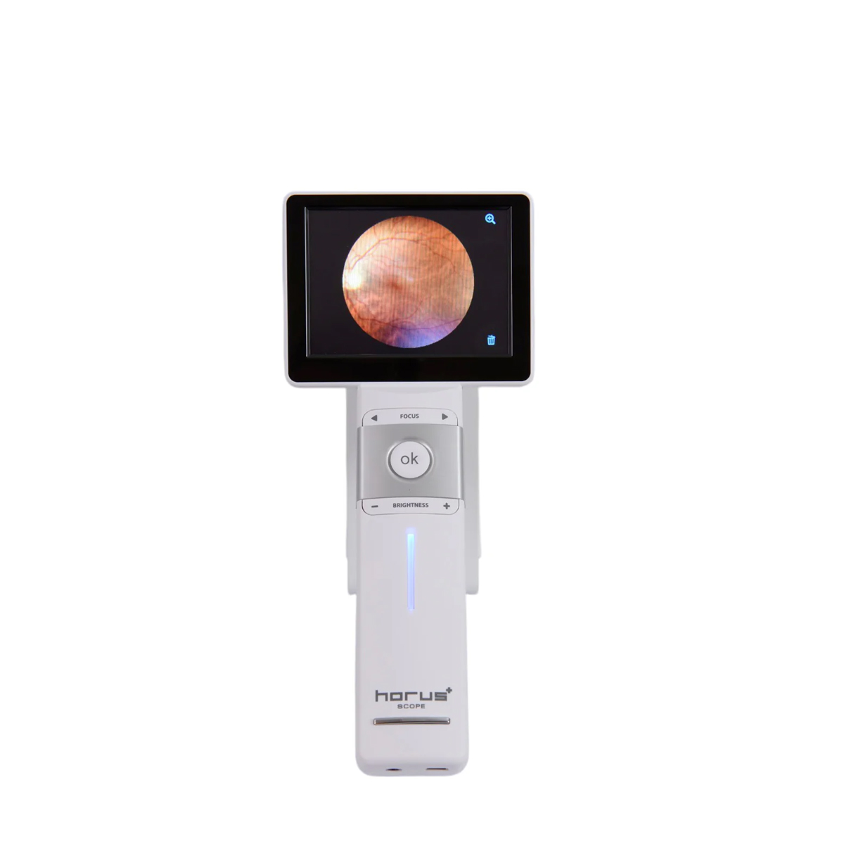
Discover the future of fundus imaging with the MiiS Fundus Camera. Compact, portable, and packed with advanced technology, the MiiS Fundus Camera combines high-resolution imaging with unmatched versatility. Effortlessly capture sharp, detailed fundus images, thanks to its 5MP sensor and 45° FOV.

Talk to a specialist

High Resolution

Captures sharp, detailed images with a 5MP sensor.

Wide Visual Field

45° field of view for precise fundus examination.

Compact Design

Portable and lightweight for use in any clinical setting.

Advanced Diagnostics

Cutting-edge technology for faster and more accurate diagnostics.

MiiS Fundus Camera (MiiS Fundus Camera)

The MiiS Fundus Camera delivers exceptional image quality, meeting ISO 10940 standards through its 5MP sensor (2592 x 1944 pixels) and a 45° field of view (FOV) for detailed fundus imaging. It features seven internal fixation targets for precise capture of the macular center, optic disc center, and peripheral areas. Designed for versatility, it offers both autofocus and manual focus (power-focus) functions for effortless image capture, a touchscreen LCD display for intuitive operation, and Wi-Fi connectivity for seamless data transfer. This innovative, portable camera is ideal for telemedicine, clinical offices, emergency departments, hospital rounds, and educational purposes, making it an indispensable tool for modern ophthalmic care.


45-Degree Field of View

Users will be impressed by the wide view offered by the MiiS Fundus Camera. Equipped with a 45-degree field of view, the camera provides a remarkably wide and detailed image of the retina, allowing healthcare professionals to observe a greater portion of the ocular structures in a single shot. This is made possible by the combination of the camera with the high-quality 45° Lens, which ensures clarity and precision.

Fixation Point

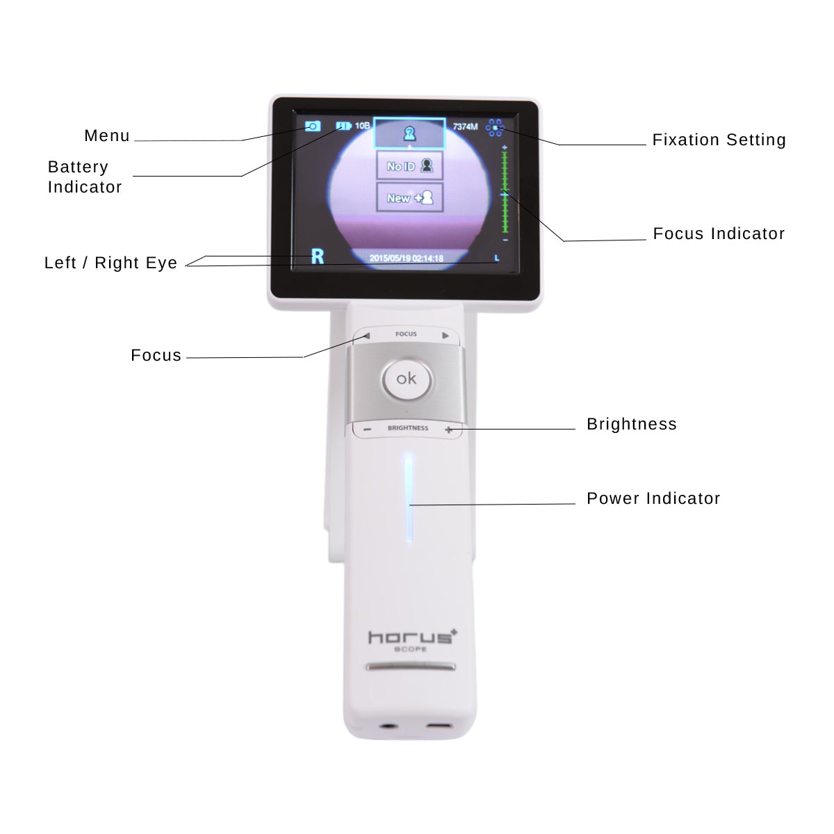
The MiiS fundus camera is equipped with 7 fixation points, providing enhanced guidance for both the clinician and the patient. These fixation points help ensure proper alignment and stability during the examination, allowing the patient to focus and maintain correct positioning. This facilitates achieving an optimal 80-degree field of view, enabling healthcare professionals to capture a wider and more detailed retinal image with greater ease. The use of multiple fixation points also contributes to improving accuracy and consistency in retinal imaging, which is essential for the diagnosis and monitoring of various ocular conditions.

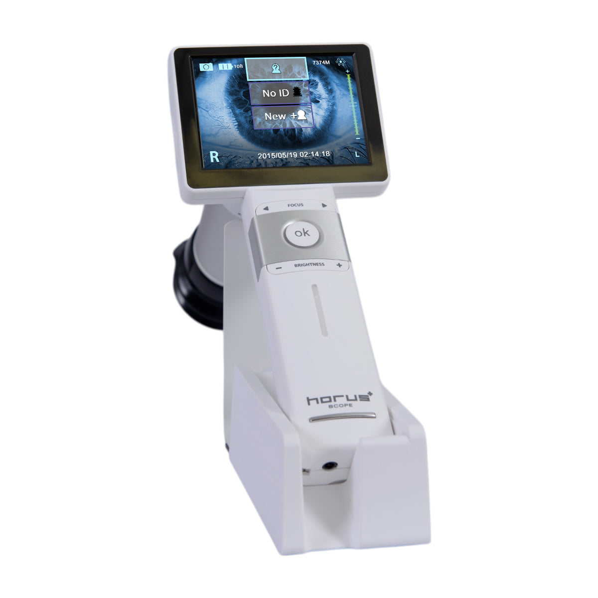
Touch Screen

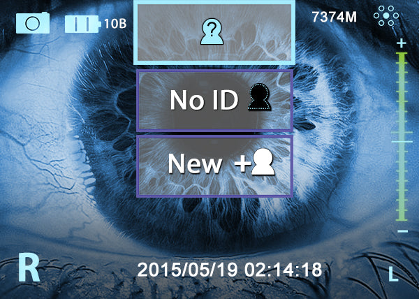
The MiiS Fundus Camera features an intuitive, high-technology touch screen that makes the device exceptionally easy to use. Designed with simplicity in mind, the touch screen interface allows healthcare professionals to effortlessly navigate the camera’s functions and settings. The responsive touch screen enhances the overall user experience, enabling quick adjustments, image capture, and result review with just a touch. This ease of use reduces training time and increases efficiency, making the MiiS Fundus Camera a practical tool for both experienced specialists and those new to retinal imaging. Its sleek design and advanced technology ensure a smooth, optimized workflow in clinical settings, enhancing both diagnostic accuracy and patient care.
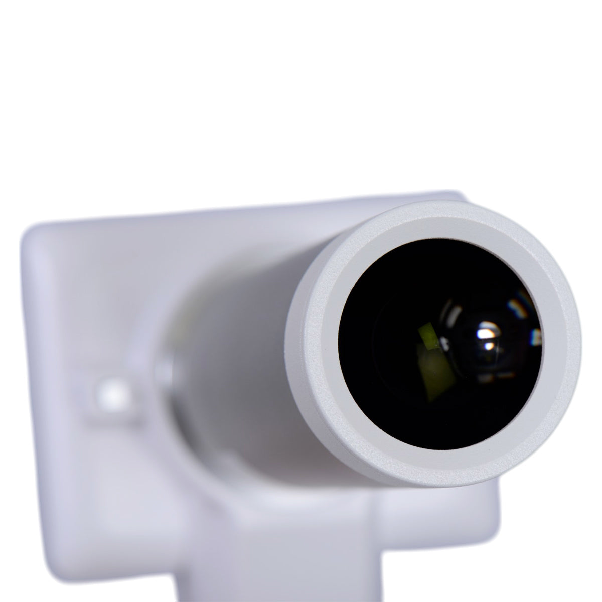
5 Megapixel Resolution

The MiiS Fundus Camera is equipped with an industry-leading 5-megapixel resolution, ensuring that every captured image is sharp, detailed, and of the highest quality. Whether in video or still image mode, the 5-megapixel resolution delivers exceptional clarity, enabling healthcare professionals to observe even the finest retinal details with remarkable precision. This level of image quality is essential for accurate diagnosis and monitoring of retinal conditions, allowing clinicians to detect subtle changes that might otherwise go unnoticed. The camera’s high-resolution capabilities also facilitate precise documentation and patient follow-up, enhancing both the effectiveness and reliability of fundus examinations.

Patient Organizer

The MiiS Fundus Camera is equipped with an intuitive and easy-to-use Patient Organizer function that facilitates the tracking and recording of patient visits. This integrated system allows healthcare professionals to quickly access patient records, document each visit, and review historical images in an organized and efficient manner. With just a few taps on the touchscreen, physicians can link retinal images to specific patient profiles, ensuring that data is secure and easily retrievable. This functionality helps improve workflow efficiency and ensures continuity of care by enabling seamless tracking of changes over time, such as the progression of retinal conditions or the effectiveness of treatments. The Patient Organizer is an invaluable tool for physicians who need to manage large volumes of patient data while maintaining accuracy and organization.

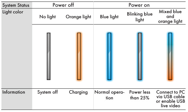
Energy Indicator

Charge while in use With the MiiS fundus camera, you will never have to worry about unexpected shutdowns. A color-coded indicator displays battery consumption and remaining life. By connecting it via mini USB, you can keep the device running while the battery charges.

Fundus examination has never been easier or more convenient than with the MiiS fundus camera, thanks to its lightweight and ergonomic design. With exceptional image quality and outstanding value, this digital fundus camera stands out as a leading choice for a wide variety of clinical settings. The MiiS fundus camera is equipped with advanced white LED and infrared (IR) illumination, ensuring optimal lighting for clear and detailed imaging under both standard and challenging conditions. Its combination of portability, high-performance technology, and cost-effectiveness makes it an ideal solution for professionals seeking a reliable and efficient retinal imaging system.

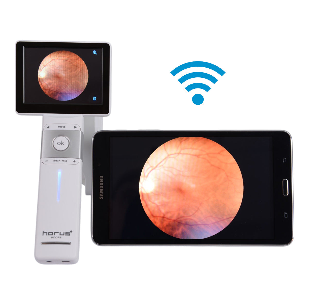
Wi-Fi Compatible

The MiiS Fundus Camera offers seamless connectivity, allowing images to be automatically transferred to any Wi-Fi-enabled computer, tablet, or smartphone. With the incorporation of EYE Fi cards, this feature enables fast and efficient image sharing, making it easier for healthcare professionals to remotely access, review, and store patient data. The wireless functionality enhances workflow efficiency by eliminating the need for manual data transfers, ensuring that retinal images are securely stored and easily accessible across multiple devices. This connectivity is ideal for telemedicine, collaborative consultations, and the overall improvement of patient care.
Auto Focus Mode

The Auto Focus method of the MiiS Fundus Camera ensures sharp and detailed images of the fundus with efficiency and precision. Leveraging advanced technology, the camera automatically adjusts the focus based on the patient’s eye position, eliminating the need for complex manual adjustments. This intelligent system evaluates in real time the ideal distance and optical plane to capture high-quality images, even in patients with slight eye movements. The Auto Focus feature not only enhances the operator’s experience by simplifying the capture process, but also ensures consistent and reliable results, essential for accurate diagnosis.
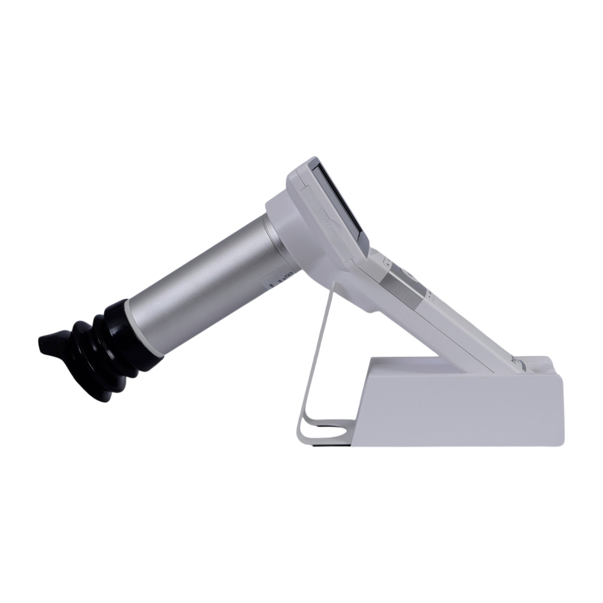
Manual Focus Mode

The Manual Focus Mode of the MiiS Fundus Camera gives operators full control over the focusing process, allowing precise adjustments tailored to the individual needs of each patient. This feature is especially useful in cases where automatic adjustments may not achieve the desired clarity, such as in patients with irregularities or challenging examination conditions. Through intuitive controls, operators can manually adjust the focus to ensure optimal image quality, guaranteeing that every detail of the fundus is captured with precision. The Manual Focus Mode is designed to complement the Auto Focus functionality, offering flexibility and adaptability for a wide variety of clinical scenarios.


Specifications

Manuals & Catalogs

Manuals

Miis Fundus Camera