









Clinical Areas:

Retina and Fundus

Glaucoma and Intraocular Pressure

Myopia Control

Cataract

  • Home
  • Huvitz HOCT-Topography Module

Huvitz HOCT-Topography Module

Comprehensive Analysis Using OCT-Based Topography: provides a Total Corneal Power Map capable of measuring the Anterior, Posterior, and Total Corneal Power Map.

Since the HOCT can analyze the posterior surface of the cornea, users can minimize errors caused by the anterior and posterior corneal axis, corneal thickness, and refractive errors produced by the vitreous and corneal structures.
Talk to a specialist

Comprehensive OCT Topography

Simultaneously maps the anterior, posterior, and total corneal surfaces.

Posterior Surface Analysis

Evaluates the posterior cornea for a comprehensive view of its structure.

Minimal Refractive Errors

Reduces errors caused by the corneal axis and vitreous structures.

Corneal Thickness Measurement

Accurately calculates corneal thickness for more reliable diagnoses.

Analysis Display / Numerical Information

Los usuarios pueden analizar con la funcionalidad de Ojo Individual, Ambos Ojos (OU) o Comparación. También es posible verificar los cambios clínicos después de un tratamiento o cirugía con la función de Progresión (Progression).

Analysis Display / Numerical Information

Users can analyze with the Single Eye, Both Eyes (OU), or Comparison functionality. It is also possible to verify clinical changes after treatment or surgery with the Progression function.

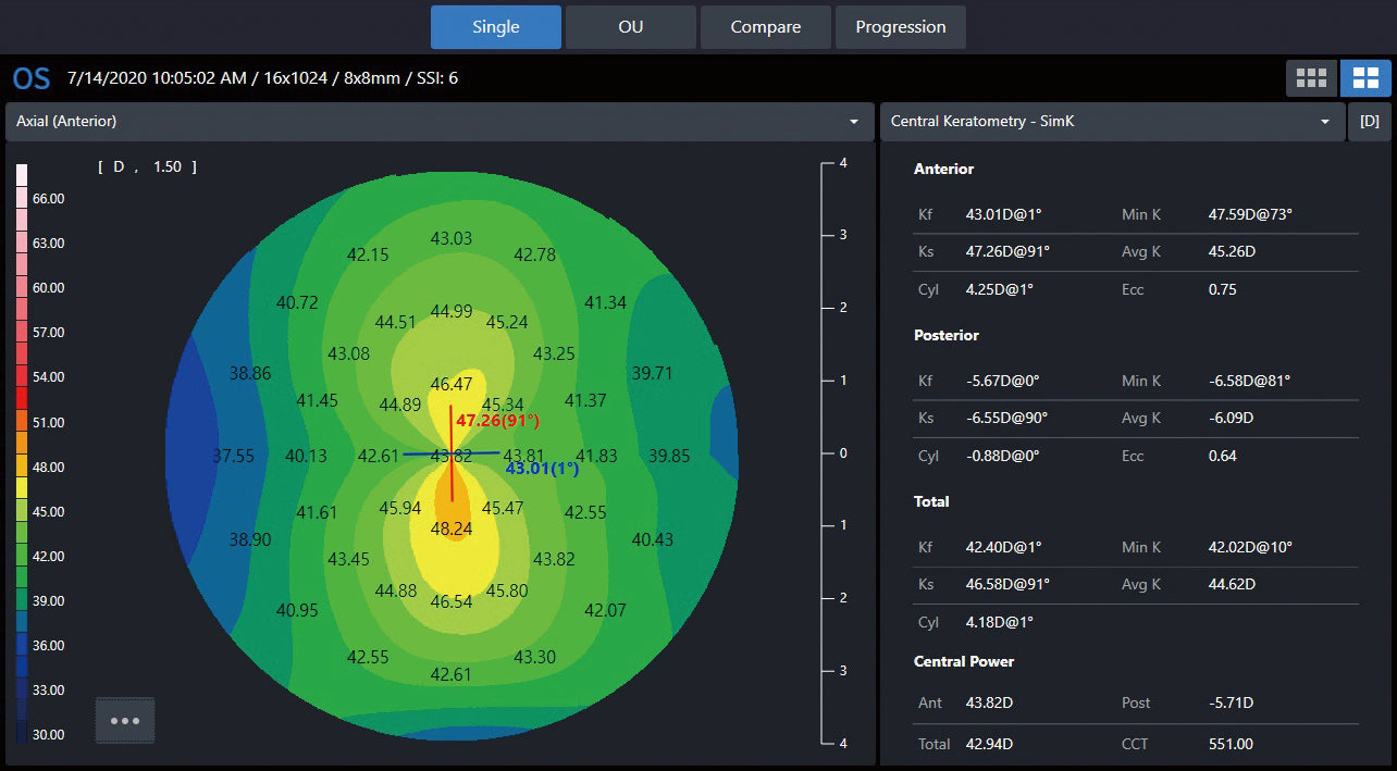
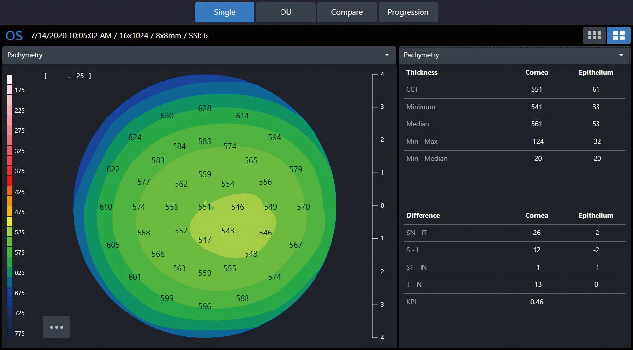
Since the map displays precise distance, size, and area with numerical information, the user can perform their analysis with confidence.

▪ Sim-K: Typical curve used for optimal IOL power calculation.
▪ Meridian: Provides the meridian of ø3, 5, 7 mm by dividing the cornea into 3 areas.
▪ Pachymetry: Total corneal thickness.
▪ Epithelium: Provides epithelial thickness at each point.


OU

COMPARISON

PROGRESSION

SIM-K


MERIDIAN


PACHYMETRY

16 Map Types for Personalized Treatment

The HOCT offers 16 map types covering both the anterior and posterior corneal surfaces. Users can visualize a wide variety of options for analysis and diagnosis. In particular, posterior corneal surface measurements enable more precise surgical outcomes than Placido or Scheimpflug methods. Motion artifacts are minimized thanks to its 2-second capture speed.