




Clinical Areas:

Retina and Fundus

Glaucoma and Intraocular Pressure

Myopia Control

Cataract

  • Home
  • Huvitz H Angio Module Angiography Module

Huvitz H Angio Module Angiography Module

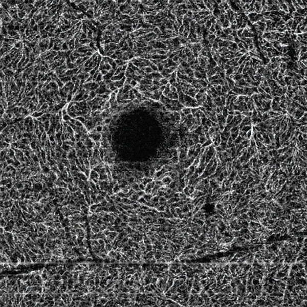
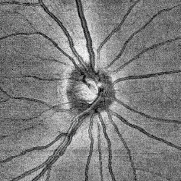
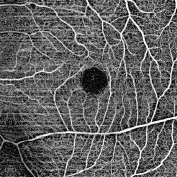
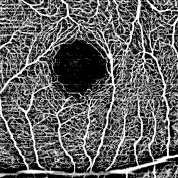
With One Single Button, precise details are obtained with high-resolution images of the retinal and choroidal vessels, as well as FAZ, flow, and density data. The Angiography module provides high-resolution images of the retinal and choroidal vessels with quantified indices. It can be used for early diagnosis and treatment monitoring of macular degeneration, diabetic retinopathy, glaucoma, hypertensive retinopathy, and retinal vein occlusion. Additionally, users can verify abnormal vasculature in the Custom View.

Since the analysis displays FAZ, Flow, and Density indices, establishing a treatment plan becomes straightforward.
Talk to a specialist

One Single Button

Get precise retina and choroid images with just the press of a button.

High Resolution

Visualize retinal and choroidal vessels with maximum quality imaging.

Early Diagnosis

Detects and monitors diseases such as glaucoma and diabetic retinopathy.

Quantified Indices

Analyzes FAZ, flow, and density to establish precise treatment plans.

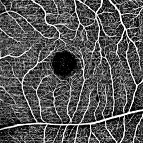
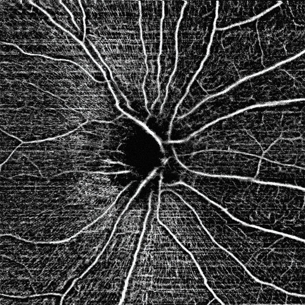
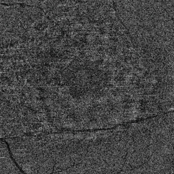
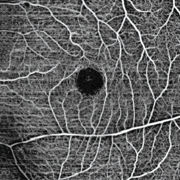
Visualization of details for precise indexing and evaluation In Detail Mode, users can specifically observe the vascular network by layer. Using the analysis tool, FAZ (Foveal Avascular Zone) details can be conveniently obtained.


6 mm SUPERFICIAL

6 mm DEEP

6 mm OUTER

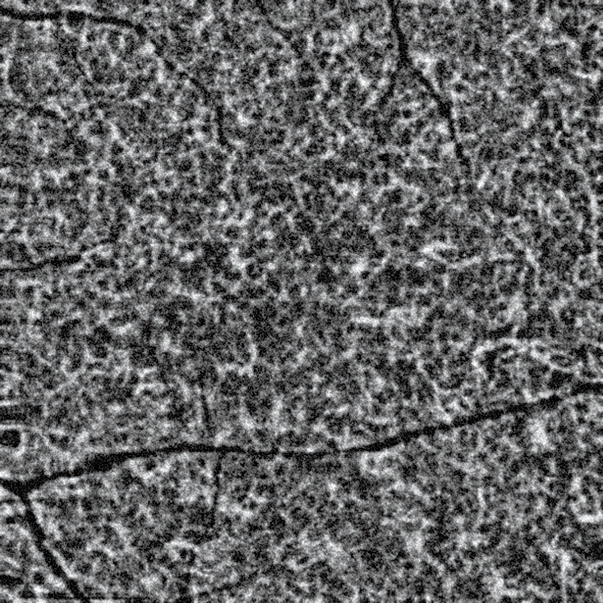
6 mm CHORIOCAPILLARIS

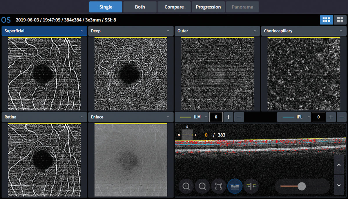
AUTOMATIC RETINAL LAYER ANALYSIS

Automatic Retinal Layer Analysis This feature provides high-resolution images of retinal and choroidal vessels with quantified indices. It can be used for early diagnosis and treatment monitoring of macular degeneration, diabetic retinopathy, glaucoma, hypertensive retinopathy, and retinal vein occlusion. Furthermore, users can verify abnormal vasculature in the Custom View. Since the analysis displays FAZ, Flows, and Density indices, establishing a treatment plan is straightforward.

Image: AUTOMATIC RETINAL LAYER ANALYSIS

AUTOMATIC FAZ DETECTION

Detailed visualization for precise indexing and evaluation In Detail Mode, users can specifically observe the vascular network by layer. Using the analysis tool, FAZ (Foveal Avascular Zone) details can be conveniently obtained.

Image: AUTOMATIC FAZ DETECTION

PROGRESSION

Progression The Progression mode helps users track the pathology of a disease.

Image: Progression

BINOCULAR COMPARISON

Binocular Comparison In Comparison Mode, users can verify the vascular network layer by layer in detail. By indicating the layers in different colors, it is easy to review and understand the pathology of a disease. In the case of comparison for diabetic retinopathy, the mode helps track the pathology and establish a treatment plan.

Imagen: BINOCULAR COMPARISON

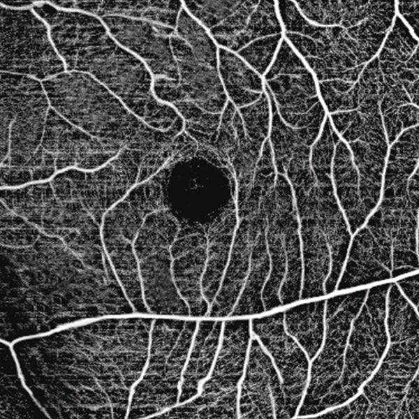
Varied Scan Sizes: 3mm / 4.5mm / 6mm / 9mm HOCT-Angiography supports a variety of scan sizes; users can choose and observe according to their needs and cases.


3MM

4.5MM

6mm

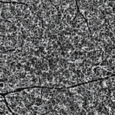
6 mm Choriocapillaris

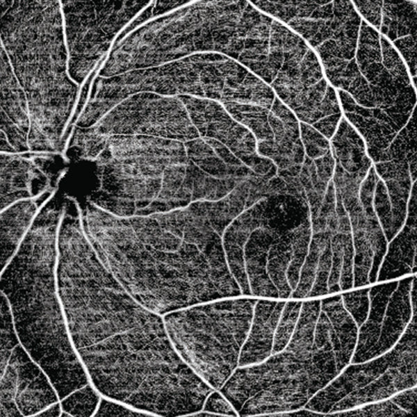
PANORAMA MODE / PANORAMIC IMAGE

Angio Panorama When reviewing a large Angiography image, it is convenient to use the Angio Panorama function. *Available modes are 3 mm (max. 12 mm x 9 mm), 4.5 mm (max. 13.5 mm x 9 mm) and manual Angio Module – Educational Video – Spanish Version

Imagen: PANORAMA MODE / PANORAMIC IMAGE