










Clinical Areas:

Retina and Fundus

Glaucoma and Intraocular Pressure

Myopia Control

Cataract

  • Home
  • Huvitz Fundus Camera HFC-1

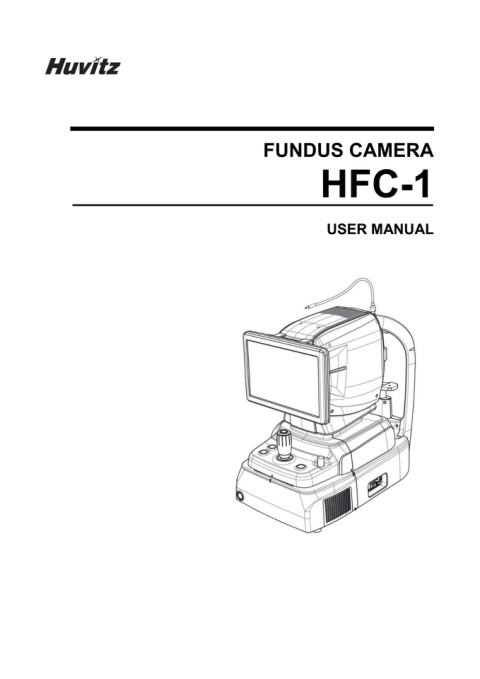
Huvitz Fundus Camera HFC-1

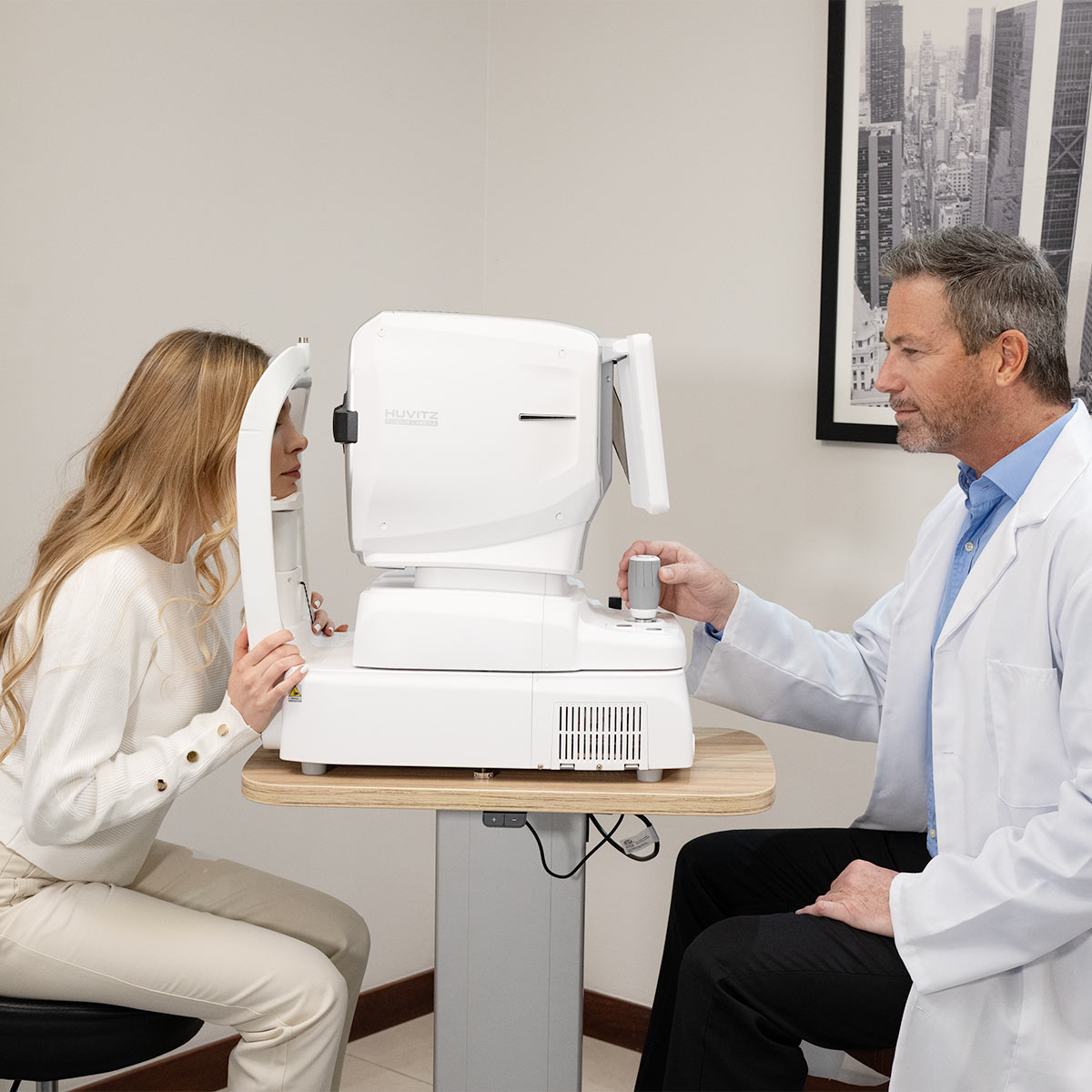
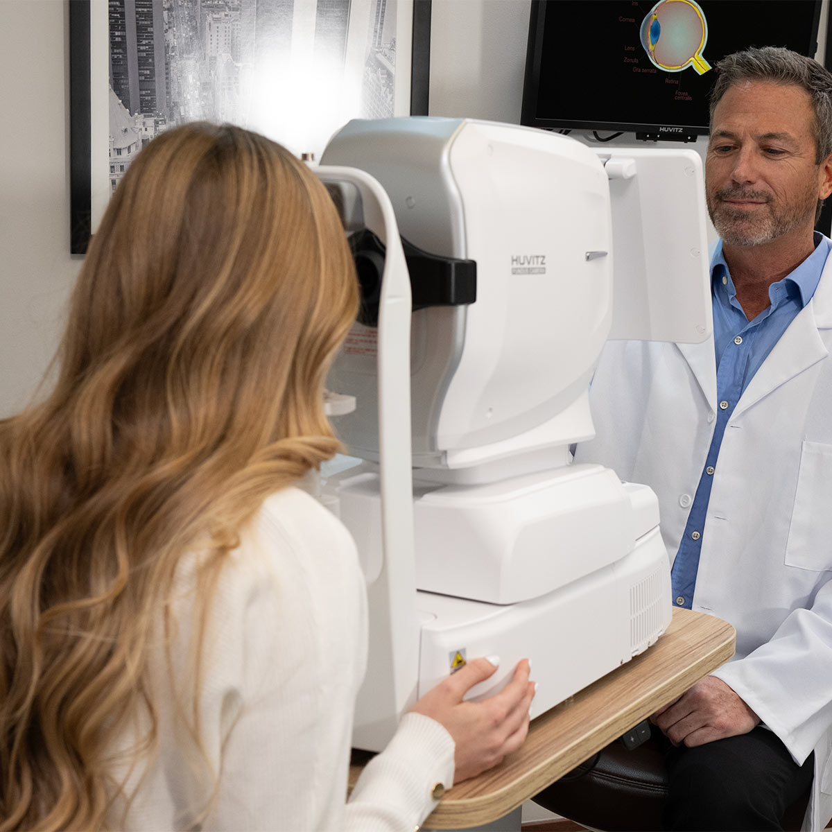
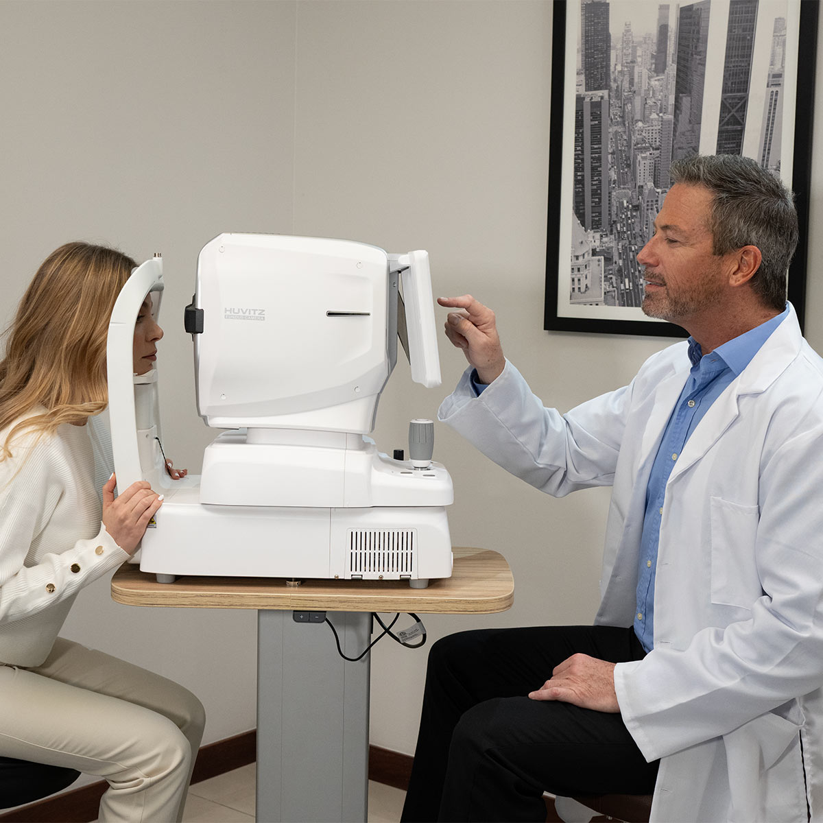
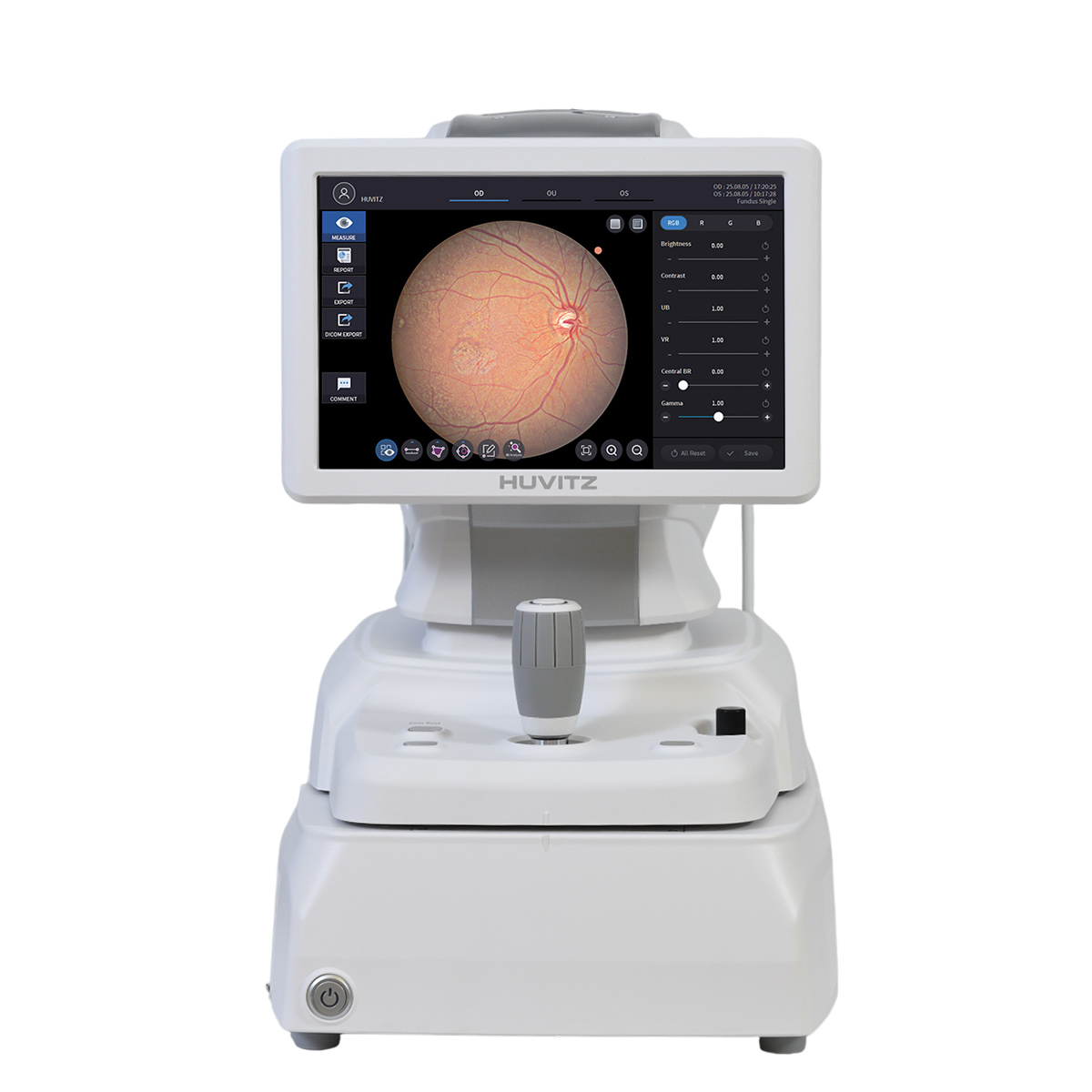
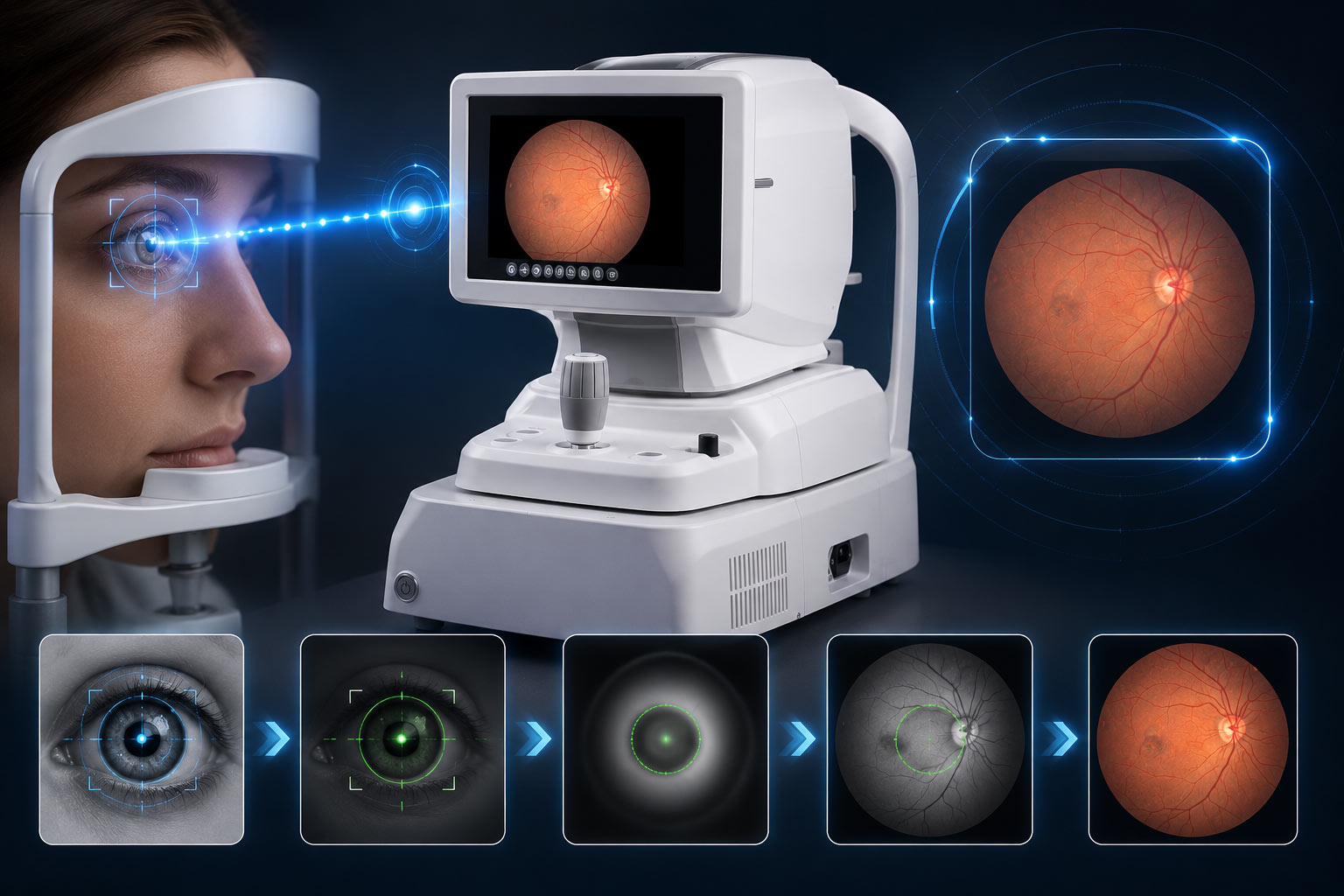
Automatic tracking and auto-shooting allow the HFC-1 Fundus Camera to adjust its modes quickly and stably on its own while measuring different pupil sizes. Its 20-megapixel high-definition camera captures images with reduced motion artifacts and features zoom capability for examining fine details.

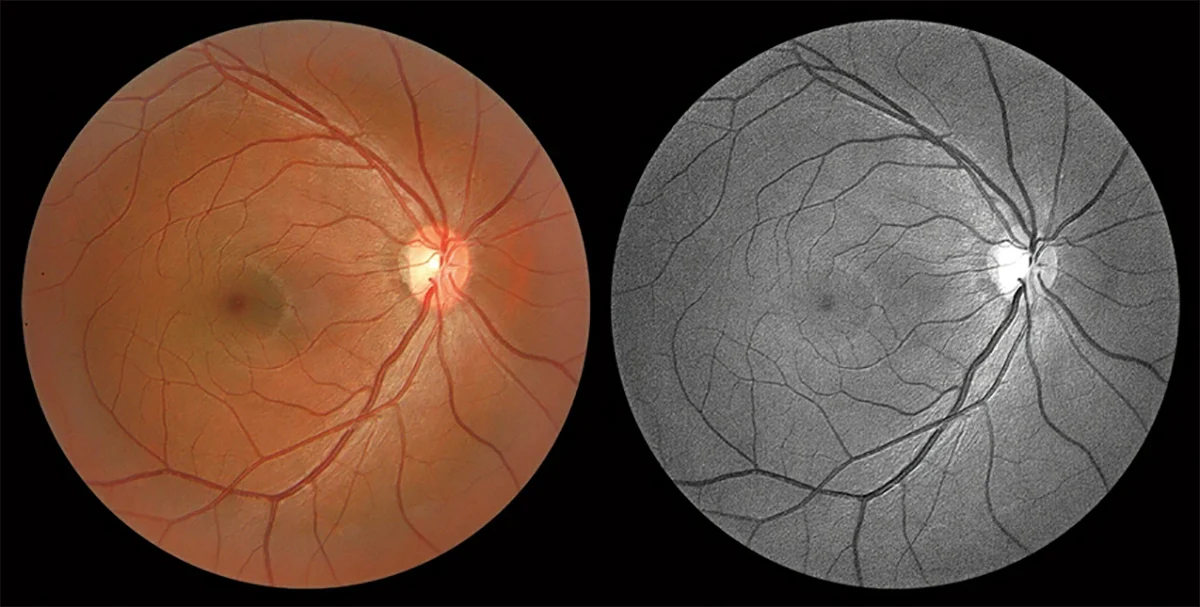
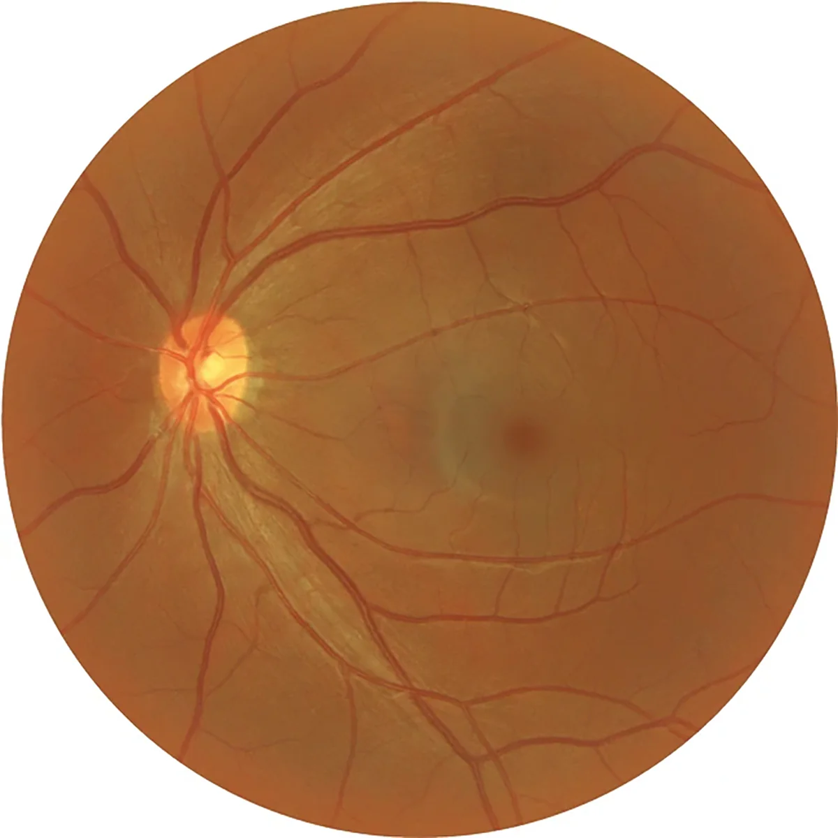
The new HFC-1 Fundus Camera includes a variety of imaging modes: color, blue, red, red-free, and cobalt, which aid in the examination of Glaucoma, RNFL, Edema, Pigmentary Abnormalities, and more.

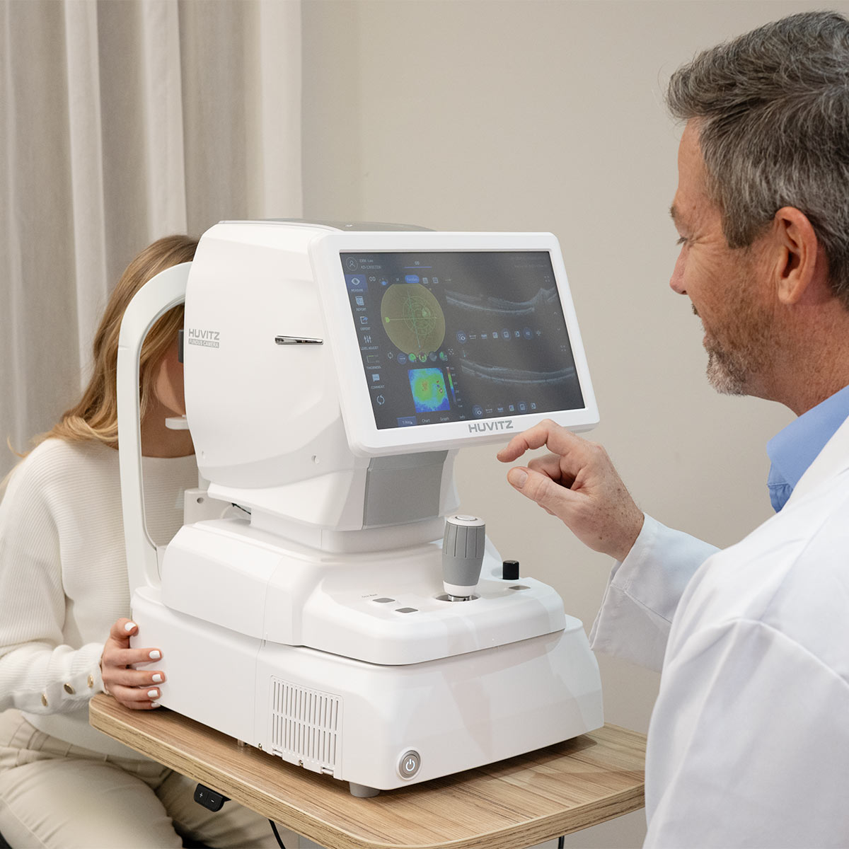
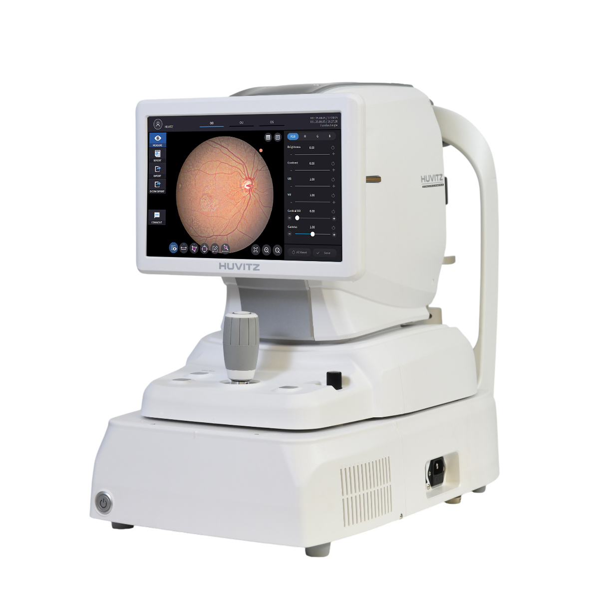
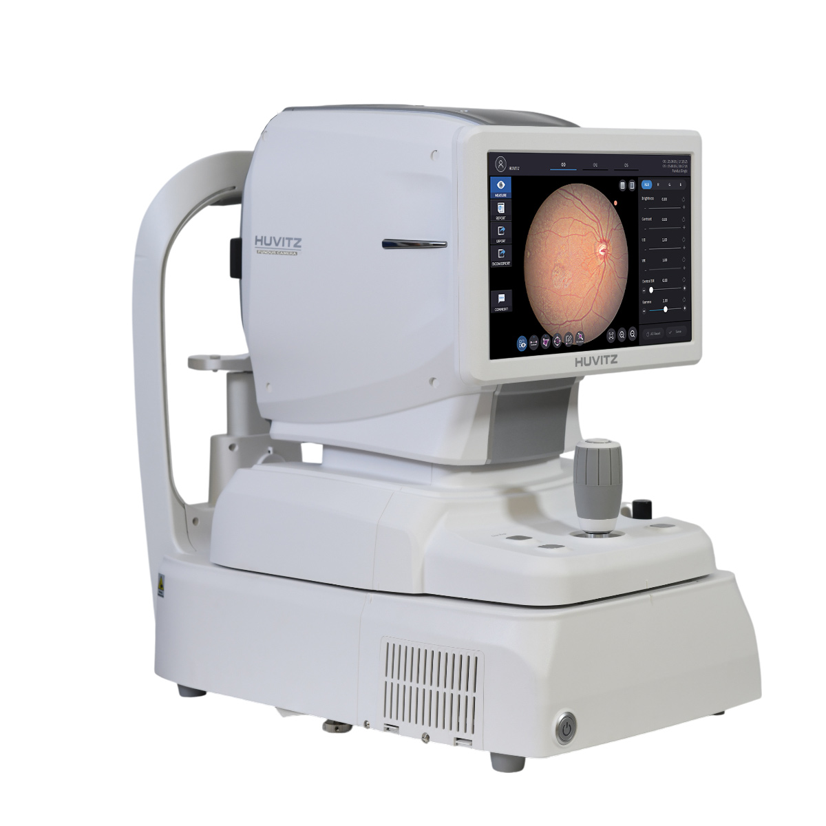
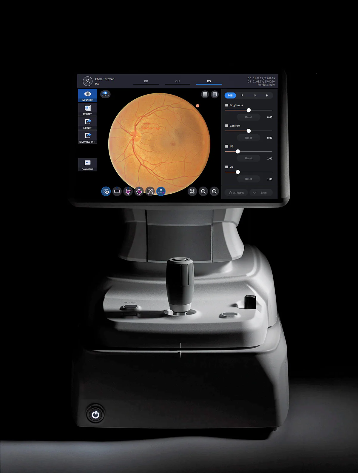
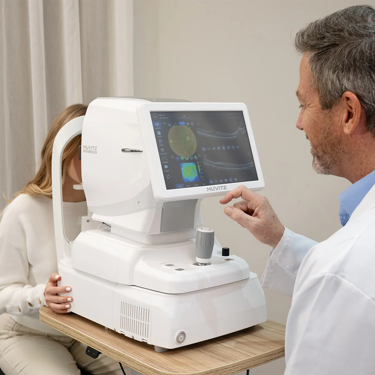
Eye care professionals can measure, analyze, diagnose, and generate reports directly from the device using the HFC-1 Fundus Camera’s built-in PC, along with its convenient 12.1″ LCD touchscreen for navigation.
Talk to a specialist

High-definition retinal imaging

Captures sharp and detailed fundus images, enabling more precise and reliable diagnoses.

Fast Capture and Simplified Operation

Optimized system enabling rapid image acquisition, reducing examination time and enhancing clinical efficiency.

Intuitive Interface and Smart Control

The modern display and simple navigation facilitate daily operation, ensuring greater productivity and ease of use.

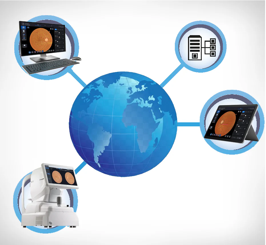
Digital Integration and Easy Sharing

Compatible with digital systems, allowing images to be stored, analyzed, and shared quickly and securely.

Featuring a large 12.1″ touchscreen and an integrated PC with 1TB of storage, the Huvitz Fundus Camera enables fast image review and analysis. By capturing detailed full-spectrum retinal images, the HFC-1 supports accurate lesion evaluation and clinical diagnosis.

Thanks to its low-intensity flash, rapid image acquisition, and Auto Tracking & Auto Shooting functions, the HFC-1 delivers excellent stability and outstanding ease of use.

User-Selectable Fundus Color Mode

Four color tone options are available for fundus images, allowing the user to choose the most suitable color mode for their diagnostic style or visual preference. This provides more precise and comfortable observation of the retinal blood vessels.

Enhanced Lesion Visibility with Adaptive Brightness and Color Control

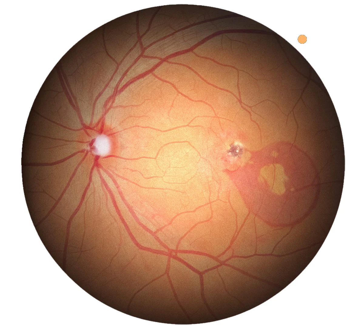
By adjusting the Central Brightness and Gamma values, it is possible to refine the brightness and color balance of the fundus image, enabling clearer visualization of areas where lesions are present. The Enhanced Visualization Technology (EVT) of the HFC-1 ensures high-quality image capture across a variety of clinical scenarios, proving especially effective in identifying subtle pathological changes.

Fixation Target

By adjusting the position of the Fixation Target on the screen, users can obtain even more precise measurement results.


Panoramic Function

This function provides essential information for the overall evaluation of the eyes, enabling the capture of high-resolution images with minimal distortion.

Auto Tracking and Auto Shooting

Fast and stable automatic tracking, along with automatic capture based on high-precision autodetection technology. There is no need to switch modes to measure small pupils, as the HFC-1 automatically adjusts the measurement process.

True-Color Fundus Scanner in a Single Shot

With 12-bit color depth and advanced gamma correction, the system captures true-color fundus images in a single shot, completely free of chromatic aberration. It delivers uniform contrast between the dark regions of the retina and the bright optic discs, enabling clear visualization of arteries, veins, and fine microvasculature. In addition, it automatically aligns and combines up to seven images to create a wide-coverage panoramic view, facilitating intuitive identification of the location and extent of lesions in a single comprehensive image.

20-Megapixel High-Resolution Camera

The efficient camera reduces motion artifacts and captures high-quality images, enabling analysis from a general overview to the magnification of fine details. Additionally, images can be viewed in multiple modes, facilitating analysis and diagnosis.

Multiple Imaging Modes:

  • Color: Bright full-spectrum images
  • Blue: RNFL, wrinkles, edema, cellular membranes
  • Red: Pigmentary changes, choroidal ruptures, nevi, melanoma
  • Red-Free: Glaucoma, diabetic retinopathy
  • Cobalt: RNFL

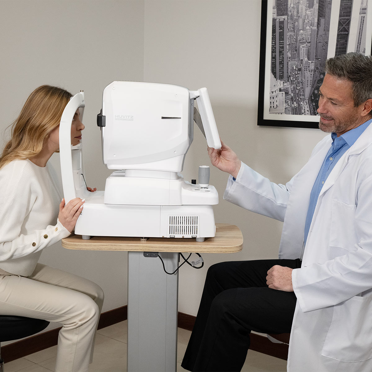
Compact Design for Space Saving

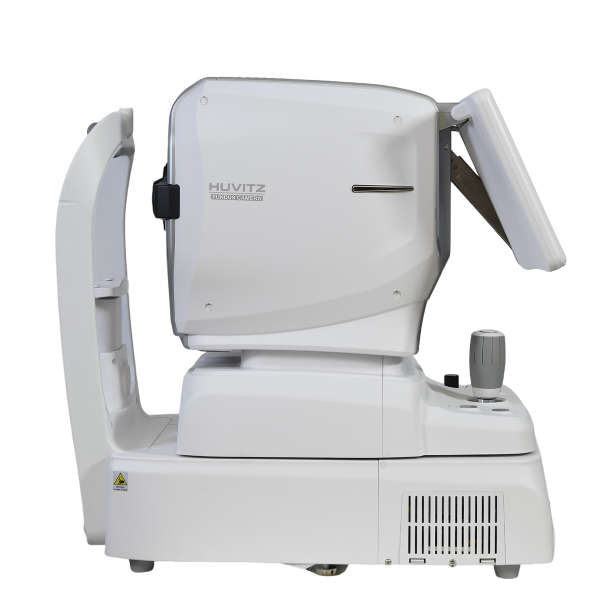
With a compact design and an integrated 1TB PC, measurements, analysis, diagnostics, and reports can all be performed in a single location. The HFC-1 was designed to optimize space usage.

12.1″ LCD Touch Screen

The HFC-1 delivers high-resolution images without ghosting thanks to its real-time image processor. With the adoption of a Wide Color TFT LCD, users can view live images with excellent quality. In addition, the touch screen significantly increases operational convenience.

Precise Comparative Analysis Based on Detailed Information

Through comprehensive and detailed analyses, professionals can clearly understand each patient’s specific symptoms, conditions, and disease progression. The system enables precise before-and-after treatment image comparison and facilitates evaluations from multiple perspectives through Single and Panorama reports, providing a deeper understanding of pathological changes.

Without the need to install additional software, users can view and analyze data directly in browsers such as Internet Explorer, Safari, and Chrome. With full DICOM compatibility, the HFC-1 remains aligned with the most advanced integration trends in medical technology.

Precise Comparative Analysis Based on Detailed Information

Through comprehensive and detailed analyses, professionals can clearly understand the specific symptoms, conditions, and progression of each patient.


Specifications

Manuals & Catalogs

Catalogs

Huvitz Fundus Camera HFC-1

Manuals

Huvitz Fundus Camera HFC-1