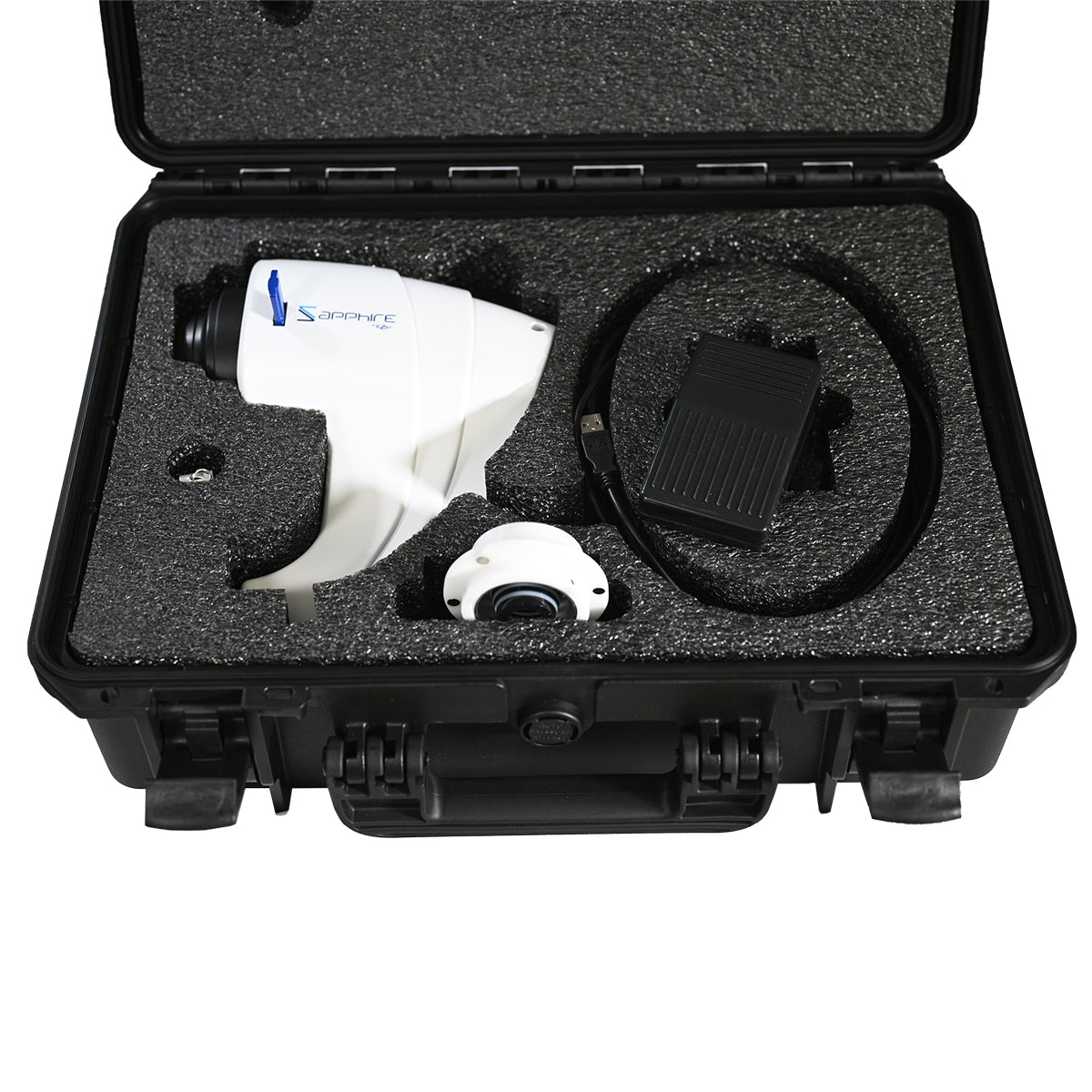
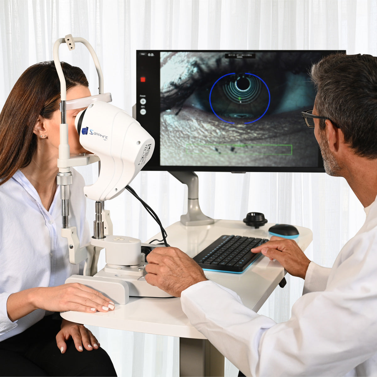
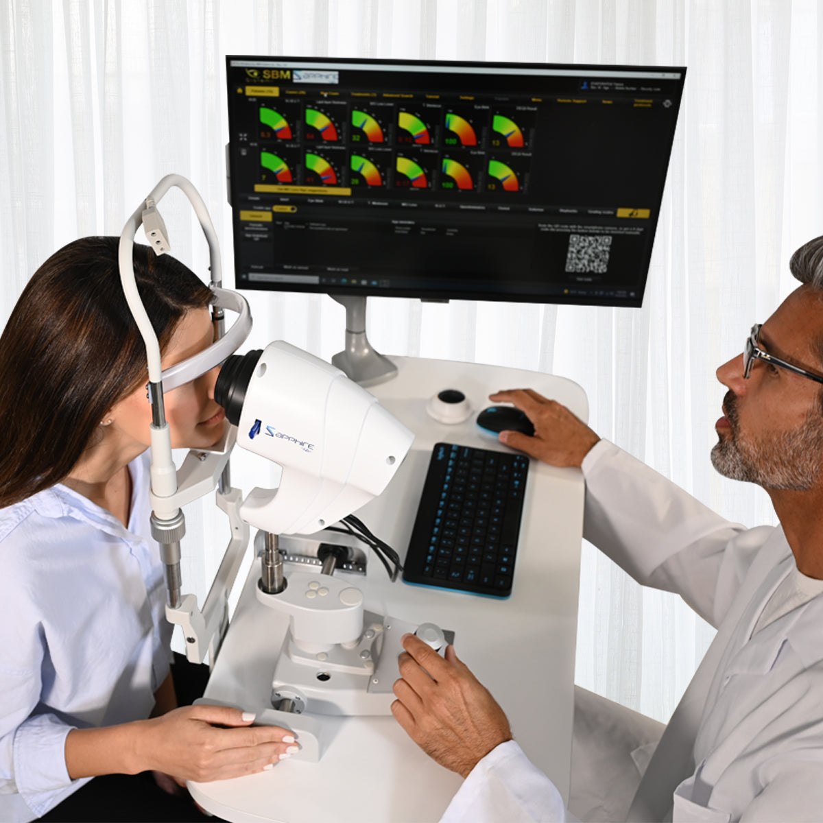
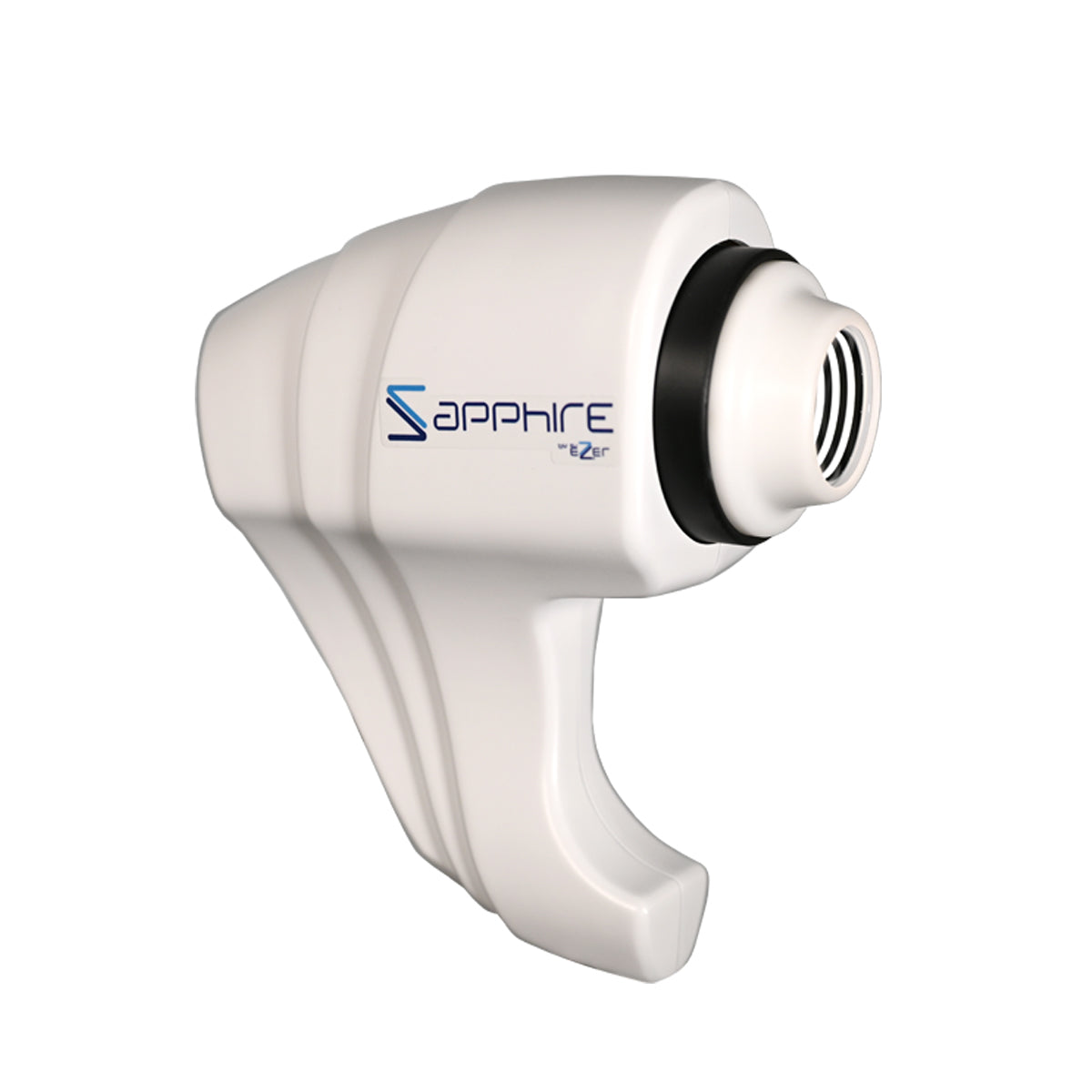
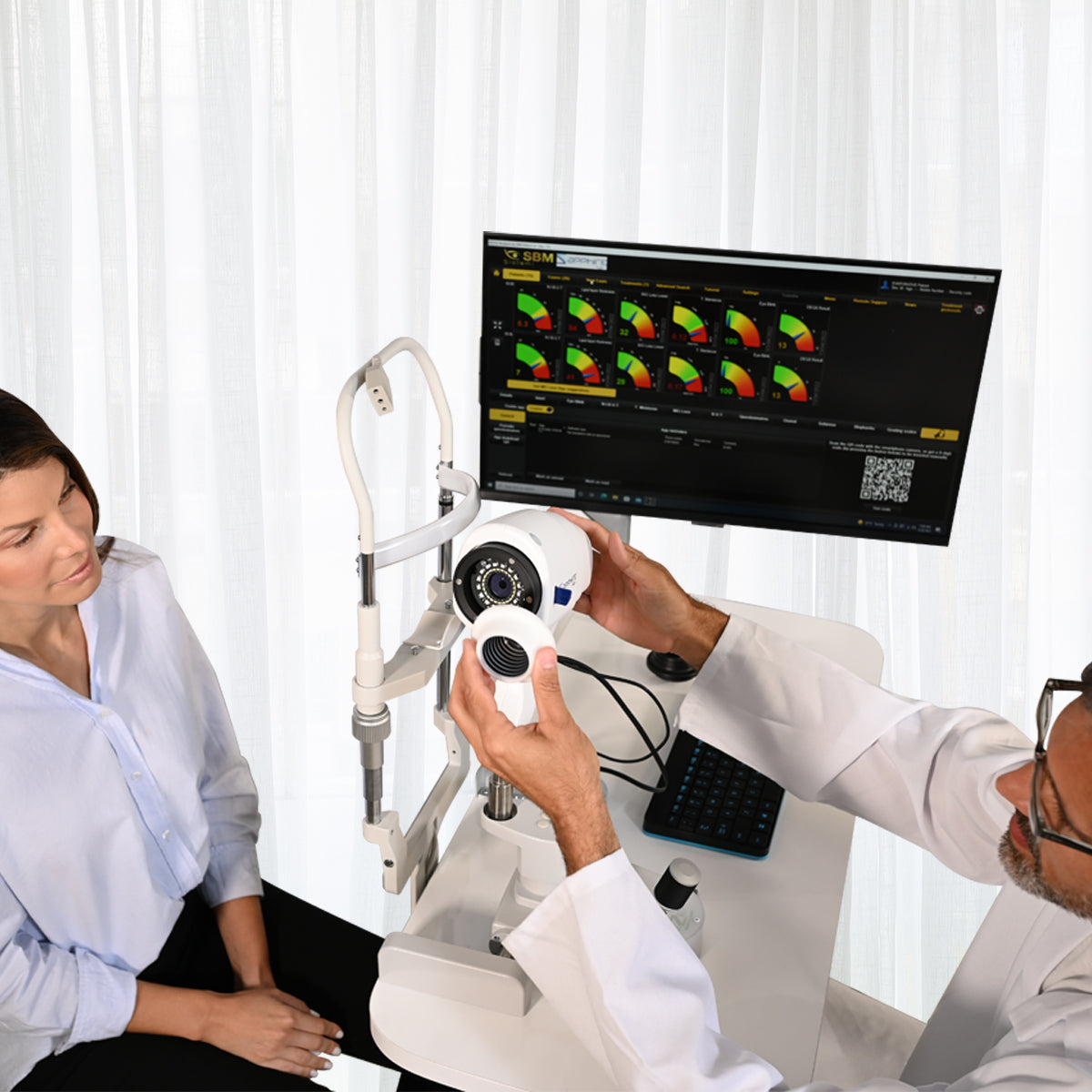
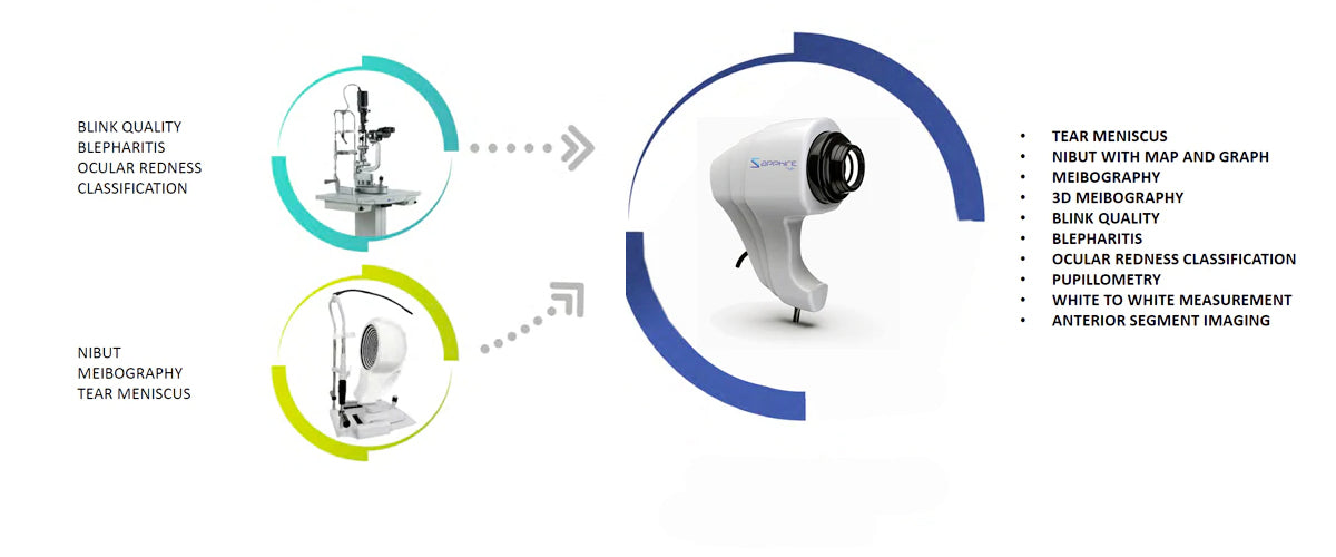
The Sapphire A device can provide you with an exceptionally detailed overview of your patient’s tear film composition. It can assist in diagnosing the type and severity of dry eye and will recommend a suggested treatment protocol for each eye individually. The device can analyze a number of different characteristics, including the lipid layer, tear meniscus, blood vessels, and more. The Sapphire A device can thus provide you with a complete picture of your patients’ ocular health, so you can determine the cause and treatment of dry eye.
Evaluates in detail the composition of the tear film for accurate dry eye diagnosis.
Personalized Diagnosis
Recommends a specific treatment protocol for each eye individually.
Multiple Ocular Parameters
Simultaneously analyzes the lipid layer, tear meniscus, blood vessels, and additional features.
Software License Included
The purchase includes a software license to take full advantage of all device functionalities.
The Sapphire A is designed to perform tear film tests, from tear quality to Meibomian Gland analysis. It is the new instrument for individual tear film analysis that allows for detailed and rapid structural investigation of tear composition.
All in One
Evaluation of all layers of the tear film
Fully Automatic Protocol
EXAM DESCRIPTION: INTERFEROMETRY
El pin ha sido construido para encajar perfectamente en el orificio que se puede ver cuando se retira la placa utilizada para el tonómetro. Cuanto más cerca esté el dispositivo del ojo, mayor será el área iluminada por el instrumento. La luz reflejada por la película lagrimal (tear film) puede observarse como un área circular blanca que cubre casi por completo la córnea.
This examination is of fundamental importance, as the majority of dry eye diseases are caused by an insufficiency in the quantity of the lipid layer. The evaluation of the lipid layer is part of your comprehensive Dry Eye assessment. Knowing what is causing Dry Eye will help determine the best treatment option.
LIPID LAYER ANALYSIS
LLT measurements are presented on an easy-to-interpret color-coded scale. Identification is performed using Dr. Guillon’s international grading scale.
EXAM DESCRIPTION: TEAR MENISCUS
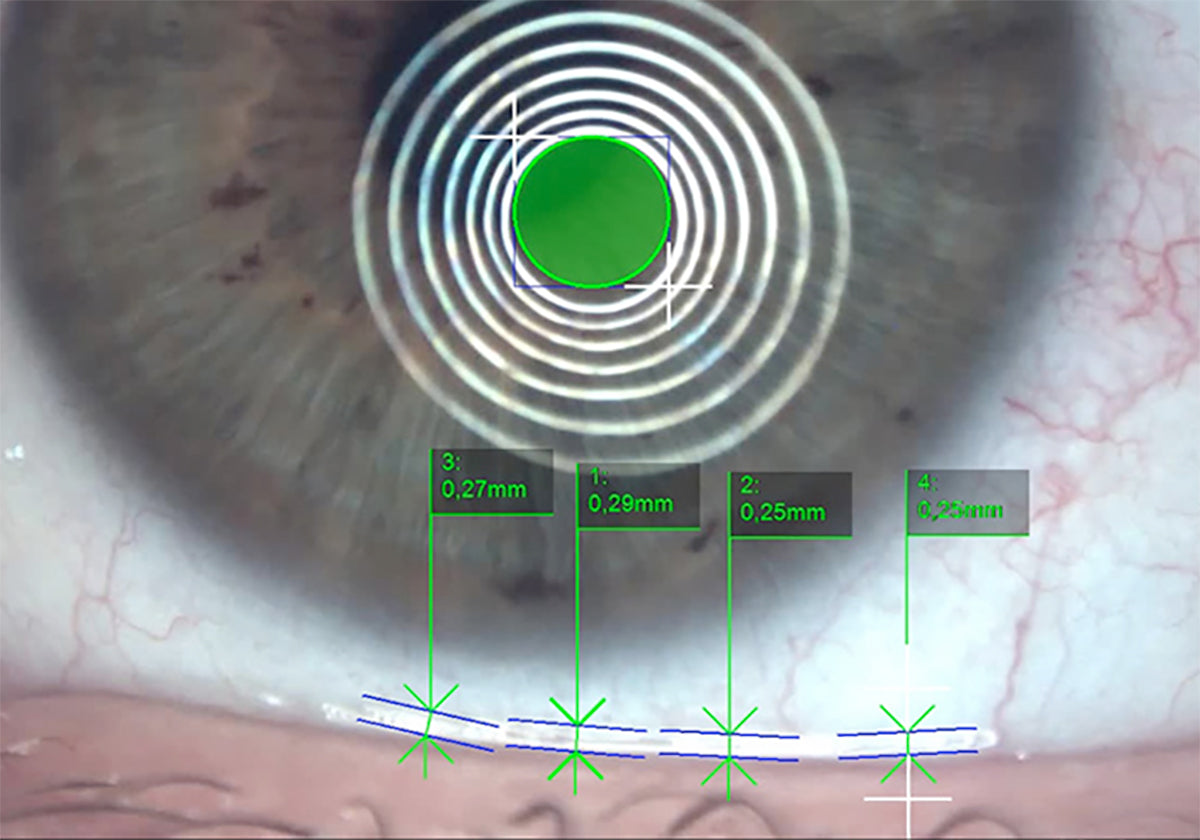
Low tear production can result in Aqueous Tear Deficiency (ATD) and cause dry eye symptoms. However, measuring tear volume is difficult, as the methods commonly used are invasive and irritating. Reflex tear production can be induced, which leads to an overestimation of basal tear flow and volume. Tear meniscus sizes are related to the tear secretion rate and tear stability, and are good indicators of total tear volume. Tear meniscus height is related to the tear secretion rate and tear stability, and for this reason is a good indicator of tear production. The aqueous layer is assessed through the non-invasive “Tear Meniscus” test and is classified into different categories. The Sapphire A device is an excellent method for screening dry eye patients, for measuring the superior and inferior tear meniscus in patients with dry eye due to Aqueous Tear Deficiency (ATD), and for determining the most effective meniscus variables in the diagnosis of dry eye. Normal tear volume is important for maintaining ocular surface physiology and ocular comfort. Total tear volume is composed of the tear meniscus, which contains between 75% and 90% of tears, the pre-ocular film, and the conjunctival fornix (cul-de-sac). Recent advances and associated software have enabled the simultaneous capture of superior and inferior meniscus images, and real-time changes have been reported.
AUTOMATED LIPID LAYER ANALYSIS
With the various magnification tools, it is possible to measure the tear meniscus height at the lower eyelid and evaluate its characteristics. The result of this examination is comparable to the Schirmer Test 1 (STT1), with the difference that it is non-invasive and lasts 3 seconds instead of several minutes.
EXAM DESCRIPTION: AUTO NIBUT
The stability of the mucin layer and the complete tear film is assessed through the study of the non-invasive breakup time (NIBUT), using the Placido cone projected onto the cornea.
The Sapphire A device allows evaluation of the stability and regularity of the tear film through non-invasive break-up time measurement (NIBUT, Non-Invasive Break-Up Time). This measurement records the number of seconds between a complete blink and the appearance of the first discontinuity in the tear film. With the Sapphire A device, from a single video, the clinician can obtain comprehensive information:
• Automatic NIBUT
• Average of more than one value
• Graph to understand the tear film stability trend throughout the video
• Tear topography showing all tear film break-up events over time.
TEAR STABILITY ASSESSMENT
By projecting the Placido disk onto the patient’s cornea, TFA automatically evaluates the stability of the tear film. The software periodically verifies the shape of the cornea using Placido rings and detects any deviation, providing break-up time values in a simple and comprehensible manner. The average BUT (AVG BUT) is presented with nomograms and, following different analyses, displays the trend line for useful follow-up after treatment.
Through the Placido rings, TFA automatically provides:
▪ First BUT
▪ Average BUT (Avg BUT)
▪ Stability graph
▪ Tear topography
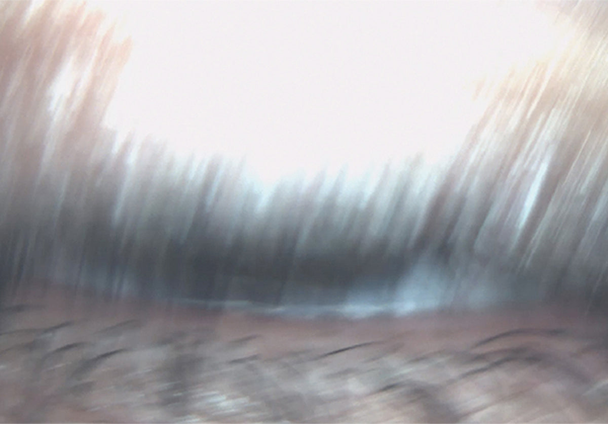
EXAMINATION DESCRIPTION: EYE BLINKING
It has been established that efficient blinking plays an important role in the health of the ocular surface, even during contact lens wear, and that it improves the performance and comfort of such lenses.
A healthy individual should exhibit periodic blinking, briefly closing the eyelids.
Most blinks are spontaneous, occurring regularly without any external stimulus. However, a reflex blink may occur in response to external stimuli such as bright light, a sudden loud noise, or an object approaching the eyes.
A voluntary or forced blink is another type of blink in which the individual deliberately closes the eyes and the lower eyelid rises to meet the upper eyelid.
A complete blink, in which the upper eyelid touches the lower eyelid, contributes to ocular surface health by providing a fresh layer of tears, as well as maintaining optical integrity through a uniform tear film over the cornea.
The blink rate and its completeness vary depending on the task performed during blink evaluation, gaze direction, the emotional state of the subjects, and the method used to measure blinking. It is also well established that the use of contact lenses (both rigid and soft) can induce significant changes in blink rate and completeness. It has been determined that efficient blinking plays an important role in ocular surface health during contact lens wear, and that it improves lens performance and comfort. Inefficient blinking during contact lens wear may be associated with a low blink rate or incomplete blinking, and can frequently be the cause of dry eye symptoms or ocular surface staining. TFA automatically detects and analyzes blinking, determining its quality.
EXAM DESCRIPTION: MEIBOGRAPHY
Meibography is the visualization of the glands by illuminating the eyelid with infrared light. It allows obtaining images of the gland morphology in order to diagnose any meibomian gland dropout that could lead to lacrimal dysfunction.
AUTOMATIC DETECTION OF MEIBOMIAN GLANDS IN UPPER AND LOWER EYELIDS
Meibomian glands (MGs) play a fundamental role in tear quality by producing lipids (meibum) that form part of the superficial tear film. MG dysfunction destabilizes tear composition, resulting in evaporative dry eye. The posterior lamella of the eyelid houses a set of parallel MGs located between the palpebral conjunctiva and the tarsal plate. A normal Meibomian gland is approximately linear and measures between 3 and 4 mm in length, traversing the posterior aspect of the eyelid perpendicular to the lid margin. A more detailed examination of a Meibomian gland reveals a tubuloacinar architecture with saccular arrangements of acini and a ductal system that communicates with orifices near the mucocutaneous junction of the eyelid. The glandular acini contain clusters of modified sebaceous cells known as meibocytes (the functional unit of the Meibomian gland). These cells synthesize and secrete lipids (meibum) into the precorneal tear film. Meibum permeates the tear surface, where it fulfills several important functions: it prevents tear evaporation and, consequently, desiccation of the ocular surface; it acts as a physical and hydrophobic barrier against the entry of environmental and organic agents; and it lubricates the ocular surface to prevent irritation, while simultaneously promoting a clear ocular image. Accordingly, tear physiology depends on the proper functioning of MGs.
THE SAPPHIRE DEVICE CAN DETECT THE LENGTH AND WIDTH OF MEIBOMIAN GLANDS VISUALIZED BY INFRARED MEIBOGRAPHY WITHOUT REQUIRING ANY USER INTERVENTION. IMAGES ARE THEN AUTOMATICALLY CLASSIFIED.
HOW IT WORKS
The system automatically analyzes images captured through a sensitive near-infrared (NIR) camera to locate the Meibomian Glands in a guided manner:
• A single examination valid for both the upper and lower eyelid;
• Automatic percentage of MG extent within the selected area
• Automatic percentage of Meibomian Gland loss area If preferred by the operator, it is also possible to manually compare the obtained images against three different related grading scales.
AUTOMATIC EYELID DETECTION
To reduce evaluation time, the software automatically detects the eyelid margin for Meibomian Gland (MG) analysis. Meibomian Gland Dysfunction (MGD) is characterized by chronic and diffuse abnormalities of the Meibomian Glands, along with alterations in meibum secretion and chemical composition. MGD leads to increased tear evaporation, elevated tear osmolarity, and greater susceptibility to ocular surface inflammation, epithelial damage, and discomfort. MGD is the leading cause of dry eye disease and affects the majority of the population. Blepharitis is a common eyelid condition that can produce symptoms ranging from burning and itching to flaking, eyelid discharge, eyelid redness, and recurrent conjunctivitis-like episodes (“pink eye”).
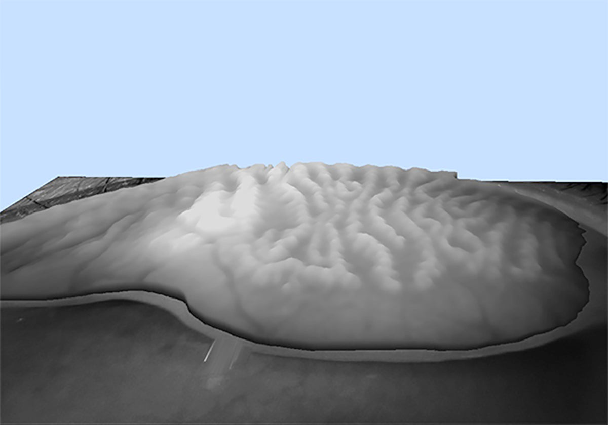
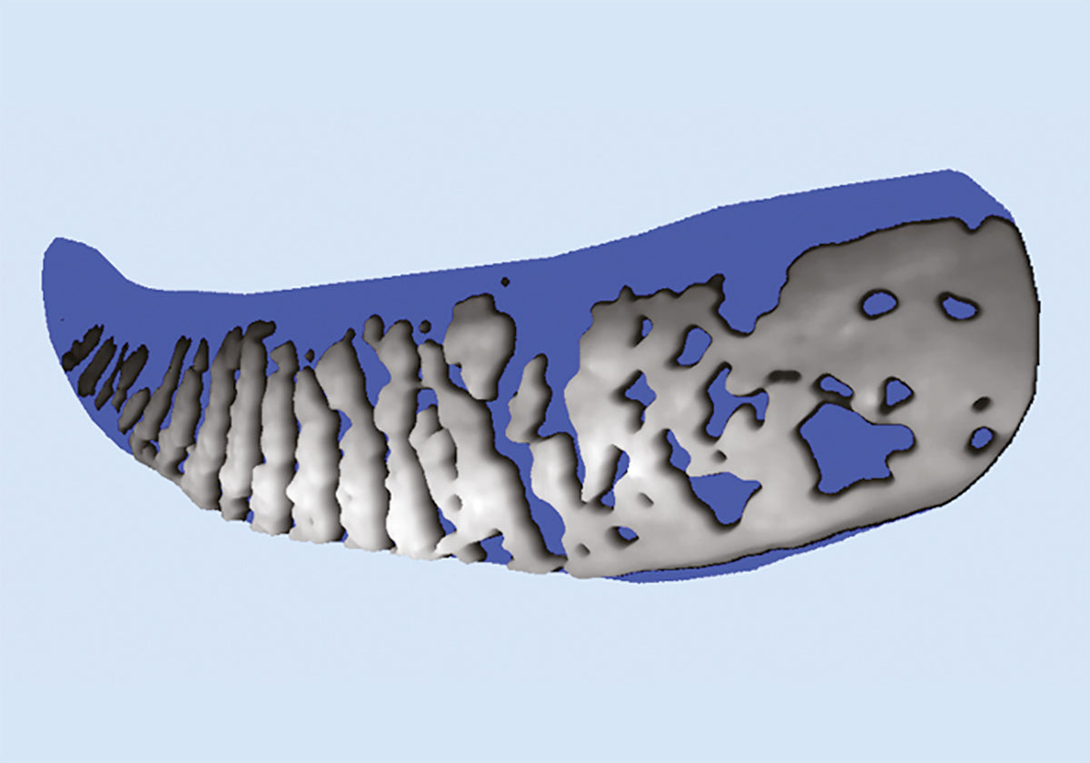
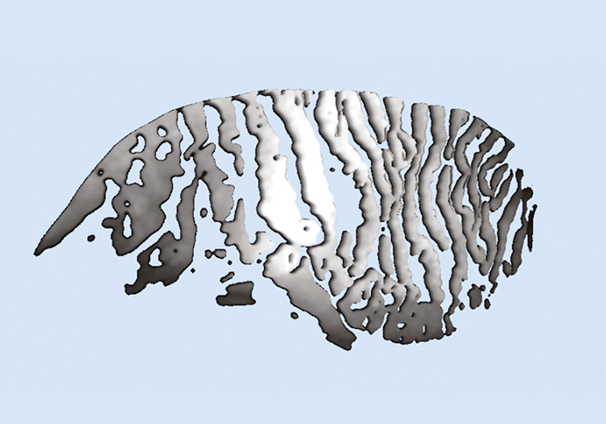
Different evaluations of the Meibomian Glands must be performed in order to prescribe the most appropriate treatment, such as Intense Pulsed Light (IPL). The Sapphire system tools provide a precise understanding of the ocular surface and, in particular, of the Meibomian Glands. The acquired images are processed and transformed into 3D images. Through scientific algorithms, the physician can visualize these 3D images, display them, and explain the anomalies to patients. This makes it easier for the physician to recommend a specific treatment, even if it involves a higher cost. It will also be possible to evaluate the efficacy of periocular Intense Pulsed Light therapy on the Meibomian Glands (MGs).
EXAM DESCRIPTION: 3D MEIBOGRAPHY
This new imaging system provides solid evidence to support the choice of a specific therapy (e.g., IPL treatment) and helps the patient understand why a particular therapy is being recommended.
AN OUTSTANDING DIAGNOSTIC EVALUATION IS NECESSARY TO DEMONSTRATE THE EFFECTIVENESS OF IPL TREATMENT TO PATIENTS
The revolutionary introduction of 3D imaging of the Meibomian Glands offers two major advantages. First, it allows confirmation of the presence of abnormal glands compared to a healthy subject through a 3D view; second, it provides a clear image to share with patients, which helps explain the possible cause of their discomfort. Furthermore, this new imaging system offers solid evidence to support the choice of a specific therapy (e.g., IPL treatment) and helps the patient understand why a particular therapy is being recommended.
PATIENT BENEFITS
• For the first time, a 3D image can help understand the structure of the eyelids. It can show possible Meibomian Gland diseases and differences compared to healthy Meibomian Glands.
• Patients can see for themselves why they experience ocular discomfort and fluctuating vision.
• Patients can understand why a specific treatment is recommended.
ADVANTAGES FOR THE PHYSICIAN
• Visualization of abnormal glandular structures in a high-resolution 3D image.
• Comparison of the glandular profile of a healthy patient with that of a patient with MGD (Meibomian Gland Dysfunction).
• Evidence supporting the diagnosis in cases of evaporative dry eye and the explanation of the reasons for choosing MGD treatment (including IPL).
• Compelling evidence to help the patient visualize what is occurring in the Meibomian glands.
• Confirmation that MGD is a contributing factor in the diagnosis of evaporative dry eye.
EXAM DESCRIPTION: BLEPHARITIS AND CYLINDRICAL DANDRUFF
This test allows direct visualization of blepharitis and the presence of Demodex. It can be performed on the external surface of the eye and eyelids.
The human skin surface harbors millions of bacteria, although some people have more than the normal and healthy amount. Blepharitis is an inflammation caused by certain bacteria found at the base of the eyelashes. These produce dandruff-like flakes on the skin, leading to infection and inflammation.
Problems with the Meibomian Glands (meibomianitis) in the eyelids can also cause blepharitis. The development of inflammation is also associated with risk factors such as dandruff, dry eye, acne rosacea, or bacteria. Blepharitis is a common ocular disorder that affects all age groups. The eye must be evaluated using a specialized instrument such as the Sapphire A magnification device. This instrument highlights inflammation in the eye and the presence of bacteria/fungi/viruses. If signs of infection are found during close monitoring, the ophthalmologist cleanses the eye and collects any discharge as a sample. This sample is subsequently evaluated under a microscope. Comprehensive Ophthalmological Examinations.
This test aids in the detection of blepharitis. It can be performed on the outer surface of the eyeball and the eyelids.
The process includes:
• Analysis of the patient’s history.
• Extrinsic detection of ocular structure, skin texture, and eyelash appearance.
• Examination of the Meibomian Gland openings, eyelash base, and eyelid margins using bright light.
• Verification of abnormalities through evaluation of tear quantity and quality.
The type of blepharitis can be determined based on the appearance of the eyelid margins. If the symptoms frequently presented by patients include slightly sticky eyelids, thickened lid margins, and absent/misdirected eyelashes, then the type of blepharitis is diagnosed as Staphylococcal.
If patients present with mild redness of the eyelids or scaling around the base of the eyelashes, seborrheic blepharitis is diagnosed. When the patient presents with obstruction of the Meibomian Glands in the eyelids, poor tear film quality, and redness of the palpebral conjunctiva, meibomian blepharitis is diagnosed. If a hard, adherent crust forms on the eyelashes and, upon its removal, small ulcers appear on the skin, ulcerative blepharitis is diagnosed. In this case, patients may experience distortion of the anterior margins of the eyelids, loss of eyelashes, and chronic tearing. In severe conditions, keratitis may also develop.
WHAT IS DEMODEX BREVIS?
Demodex brevis is a type of mite found on human skin. Like its counterpart Demodex folliculorum, D. brevis occurs naturally. D. brevis is so small that the mites cannot be observed macroscopically.
EXAMINATION DESCRIPTION: SUGGESTIONS BY DR. LUCA VIGO
Suggestions for diagnosis and treatment based on the clinical procedure of Dr. Luca Vigo and Studio Carones (Milan, Italy).
A comprehensive database focused on dry eye allows for a correct understanding and diagnosis of dry eye patients. With the practical results tab, the ophthalmologist can review the complete tear film evaluation, identifying all deficiencies causing the pathology while simultaneously understanding what treatment is needed to address each case.
DIAGNOSTIC SUGGESTION
Ocular surface data and pathology classification
Thanks to the expertise of Studio Medico Carones with Dr. Luca Vigo, TFA includes a suggestion algorithm capable of sharing a possible treatment approach for each patient. All suggestions may be useful for diagnosis and treatment.
TREATMENT MANAGEMENT
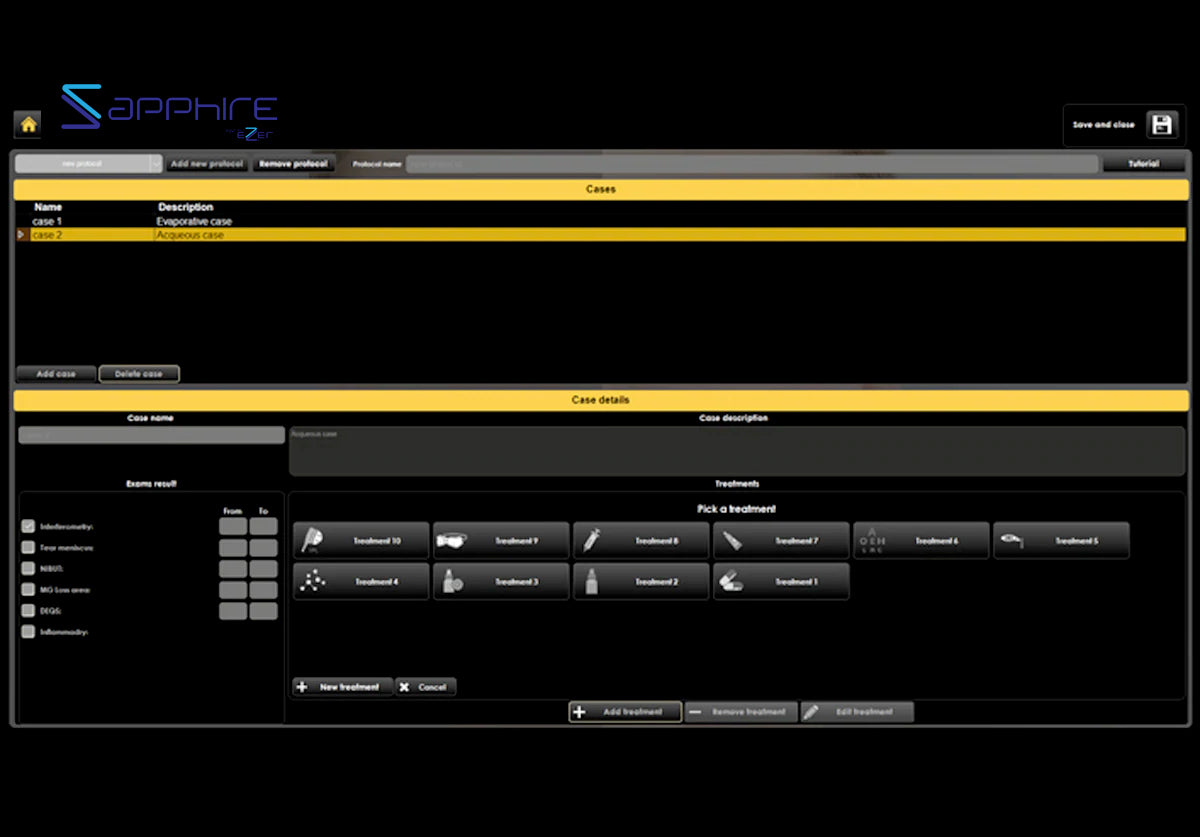
Through TREATMENT MANAGING tab, the software allows the physician to fill in the database with all drugs, integrators and treatmenrs available in his practice.
Any treatments as vitamins, Omega-3, eye drops, hot packs and IPL/Radiofrequency, can be loaded on the software to prescribe the products of the brands that the doctor prefers.
The whole report with the diagnosis and treatment suggested by the ophthalmologist will be printed directly.
PROTOCOL
(MD L.VIGO Studio Carones Milano) is already included and suggests treatment based on our experience with key opinion leaders (KOL).
All users can customize their own protocol by adding treatment procedures to be automatically selected right after the examinations are performed (this also allows diagnosis to be delegated to an assistant).
OTHER POSSIBLE EXAMINATIONS
AN EVALUATION OF GRADING SCALES FOR MEIBOGRAPHY IMAGES
The evaluation of Meibomian Gland dysfunction is of growing interest in research and clinical practice. Consequently, the assessment of MG morphology through Meibography is of great interest to both researchers and clinicians.
WHITE-TO-WHITE MEASUREMENT
Evaluation of the corneal diameter from limbus to limbus (white-to-white distance, WTW).
BULBAR HYPEREMIA CLASSIFICATION
By acquiring an image of the conjunctiva, it will be possible to compare the patient’s condition with different international classification scales.
PUPILLOMETRY
Pupillary diameter measurement has gained increasing importance in the field of refractive surgery. Larger scotopic pupil sizes may be partially responsible for the appearance of postoperative symptoms such as halos, glare, and monocular diplopia. Refractive surgeons also require accurate scotopic pupillary measurement to determine appropriate treatment zones in excimer laser surgery, corneal surgery, and intraocular surgery.
COMPARISON WITH THE MAIN INTERNATIONAL GRADING SCALES
The TFA software is a platform dedicated to dry eye that allows, in addition to assisting in the diagnosis and classification of diseases, printing and saving various medical reports, offering the most professional and clinical solutions for patients. For customer satisfaction, it is often advisable to provide technical documentation related to the examinations performed. Thanks to the various print reports of the Sapphire A device, you will have the possibility to visually explain and simply demonstrate the status of the pathology. Furthermore, it is possible to explain how the pathology has evolved over time.
COMPLETE REPORT
Complete report with all results and images used to explain to the patient any dry eye category.
TREATMENT REPORT
Patient-oriented report explaining the causes of the pathology and the recommended treatments.
FOLLOW-UP REPORT
For each value it is possible to display the trend line before/during/after treatment.
DAILY REPORT
Brief single-page report to display all examination results at a glance.
MONOCULAR REPORT
For saving and printing a single exam of interest.
BINOCULAR REPORT
For saving both eyes’ examination in a single pdf file.
The Sapphire A device provides 3D imaging to help you explain the cause of dry eye and support your treatment recommendations. It gives you the ideal tool to easily explain the source of each patient’s ocular discomfort, while helping every patient understand the rationale behind your recommendation. The Sapphire A device allows you to offer peace of mind to your patients, with the assurance that you are using the most advanced technology for accurate diagnosis and treatment.
Specifications
Image Resolution
5 MP
Acquisition mode
Multi-shot, video
Focus
Autofocus, manual focus
ISO Management
Variable
Cones
Main cone and Placido cone
Camera
Color, near-infrared sensitive (NIR), with yellow filter