









Clinical Areas:

Retina and Fundus

Glaucoma and Intraocular Pressure

Myopia Control

Cataract

  • Home
  • Huvitz Optical Coherence Tomographer with Fundus HOCT-1F (80)

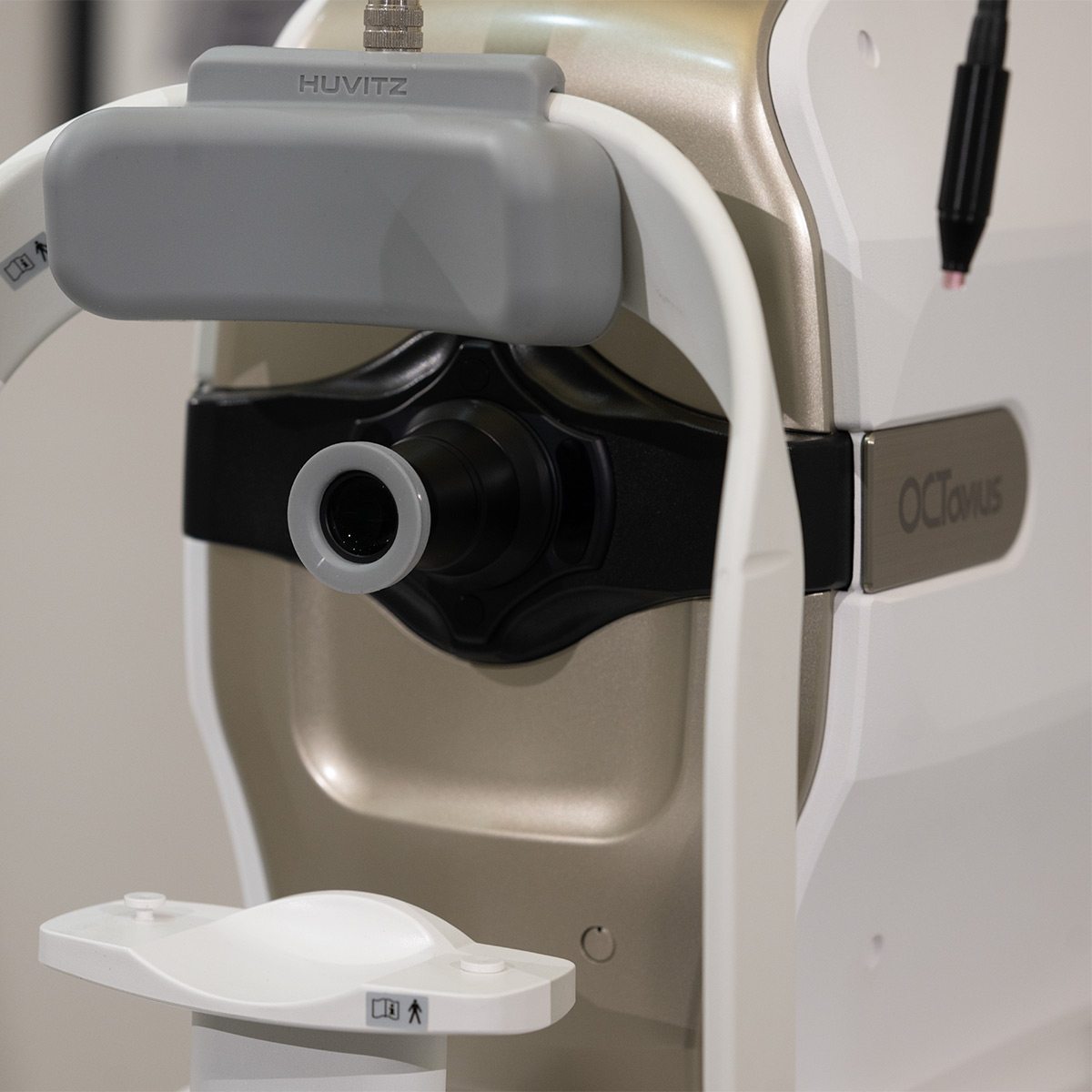
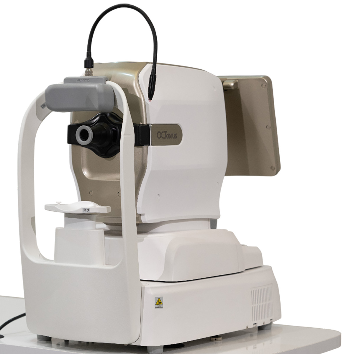
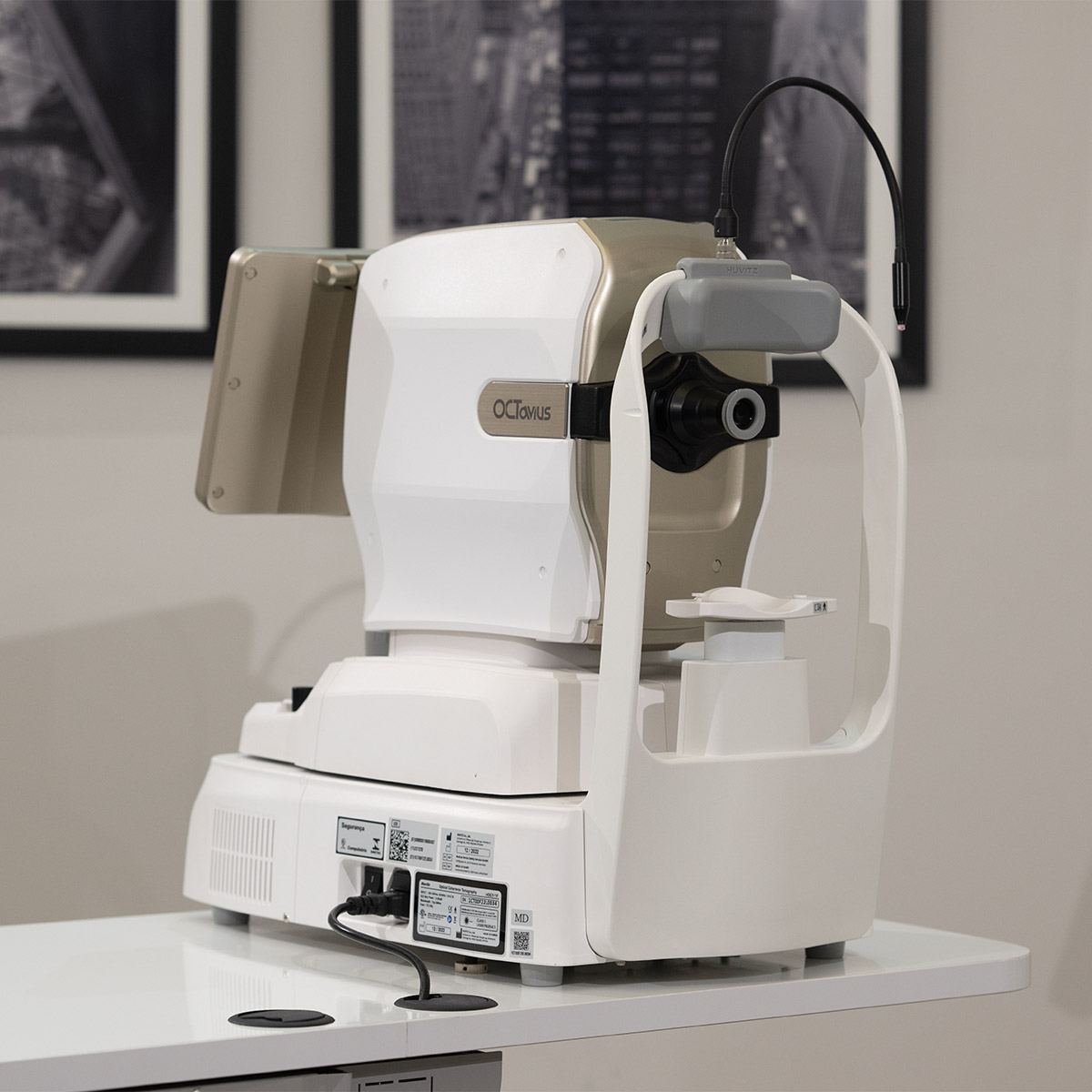
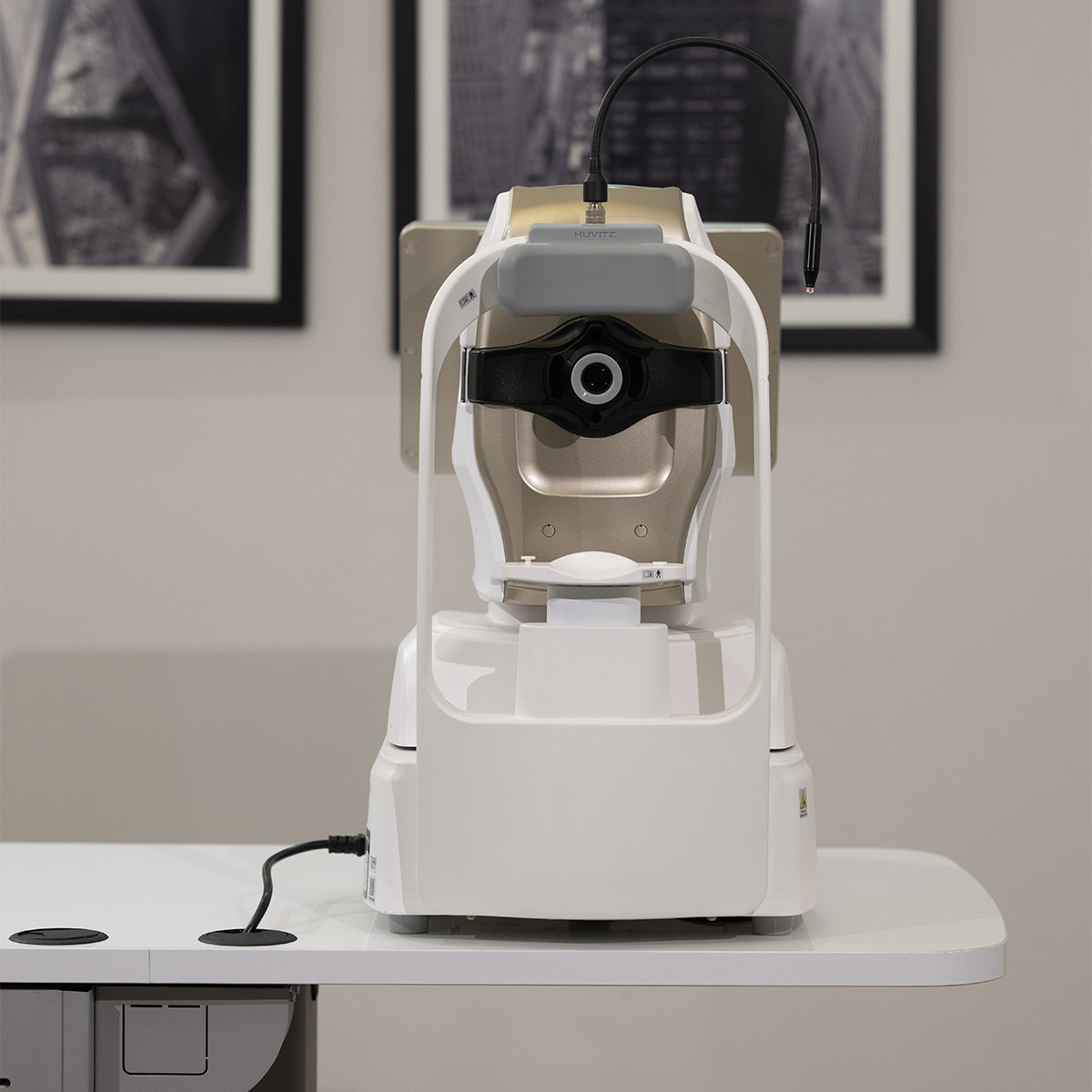
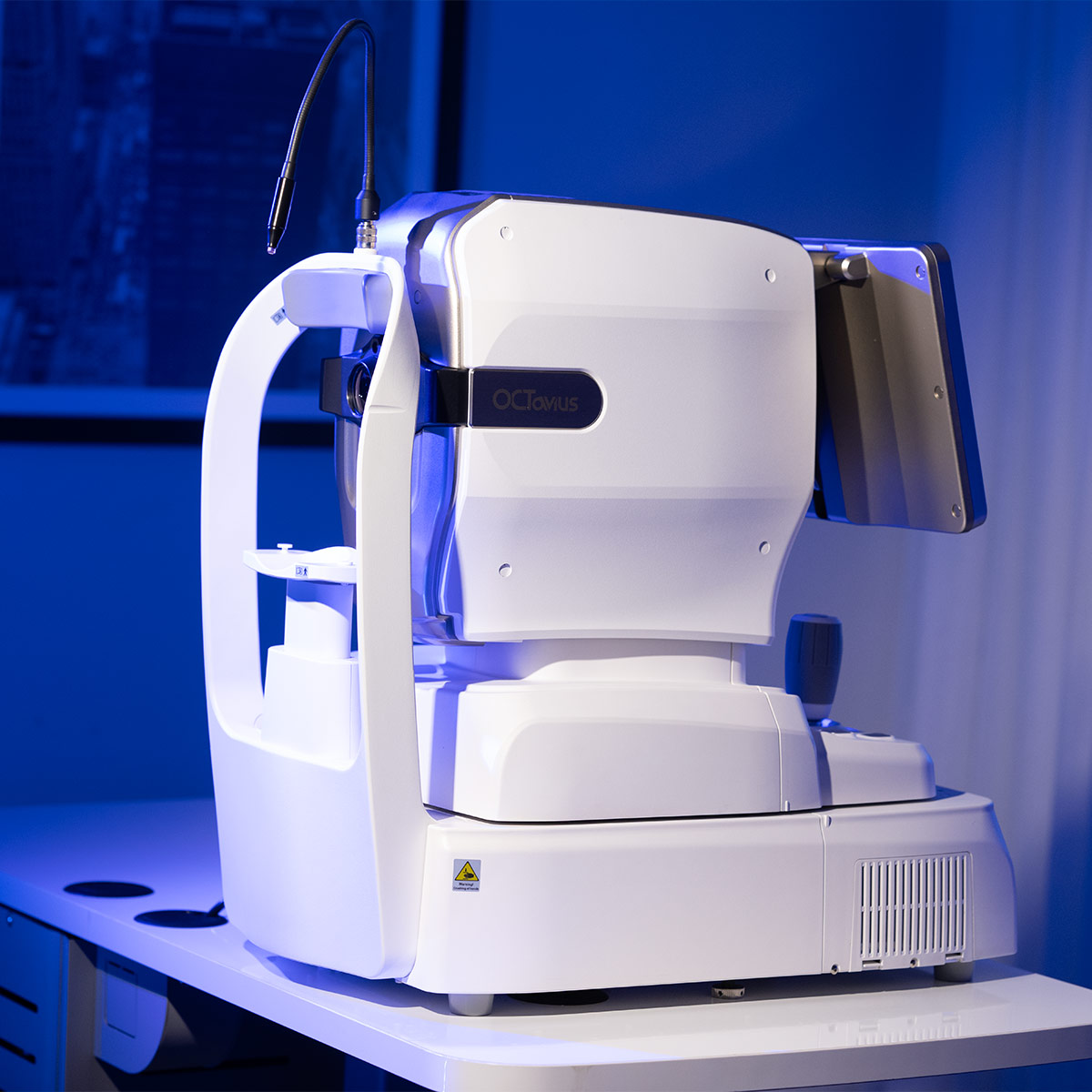
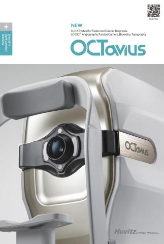
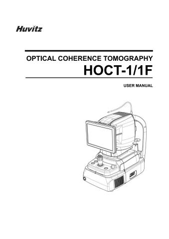
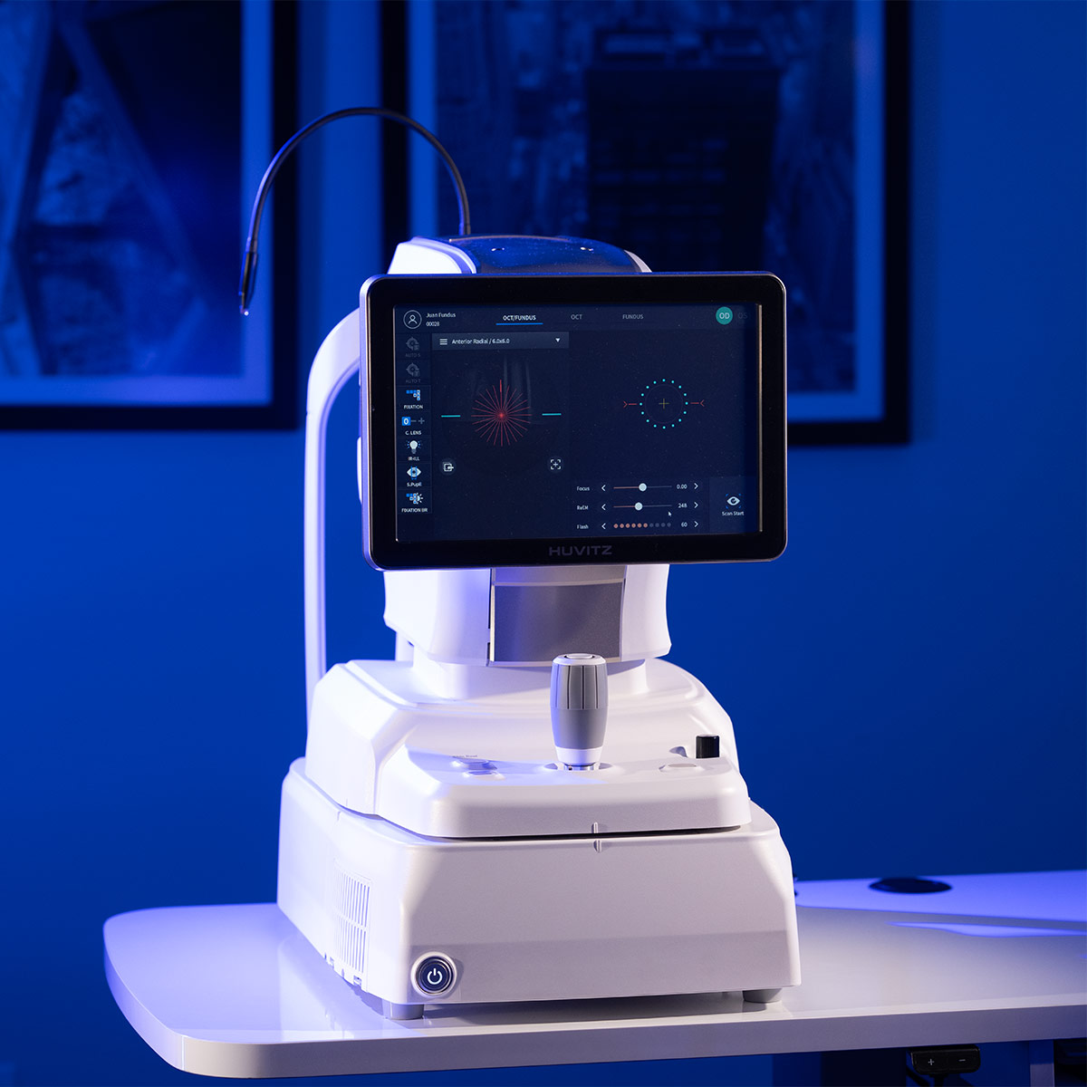
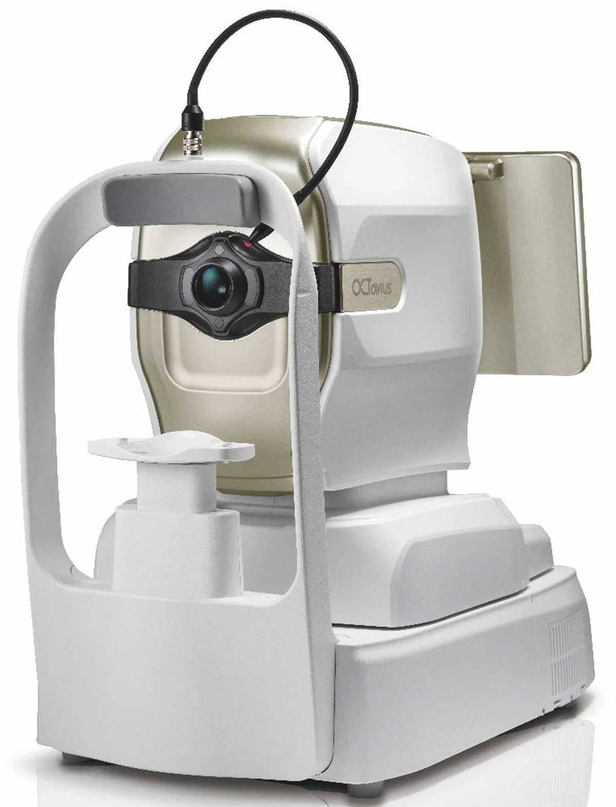
Huvitz Optical Coherence Tomographer with Fundus HOCT-1F (80)

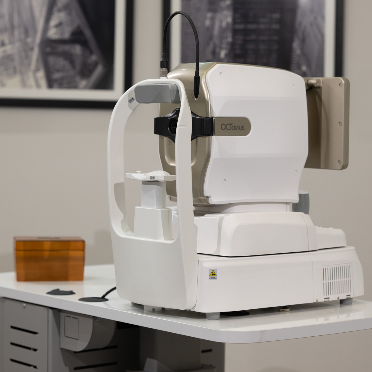
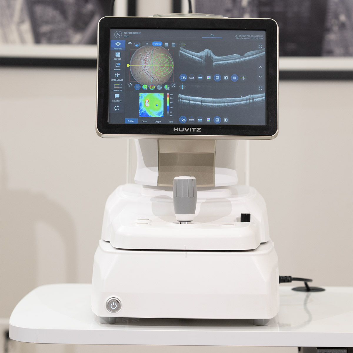
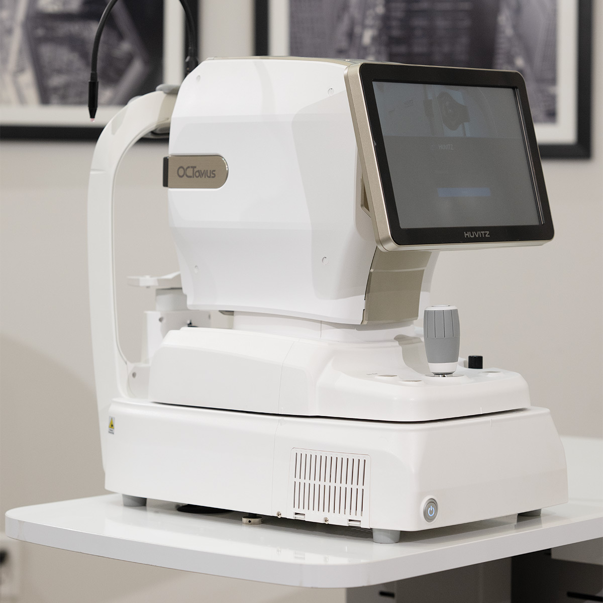
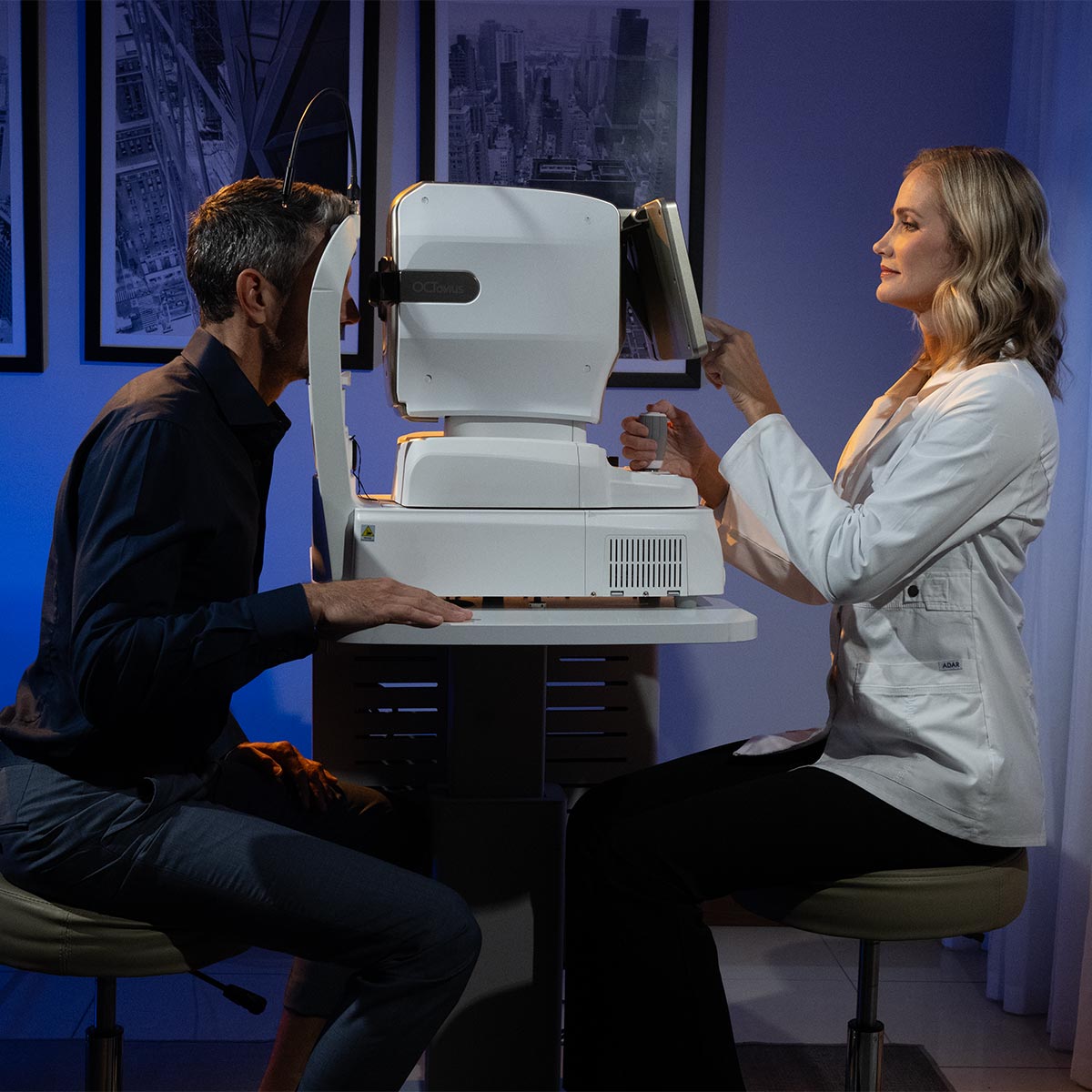
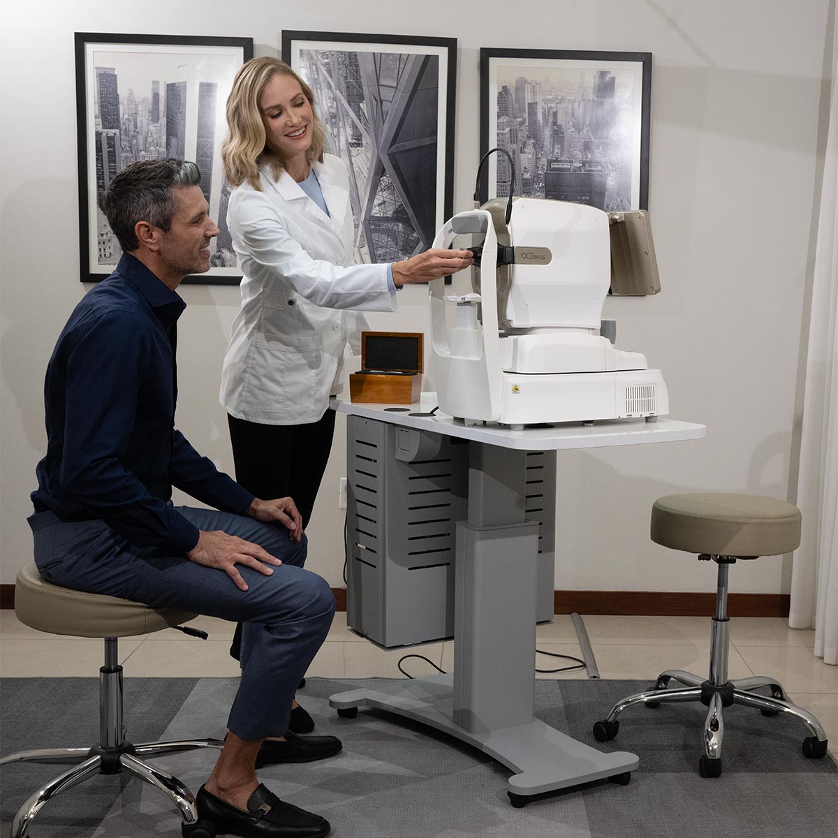
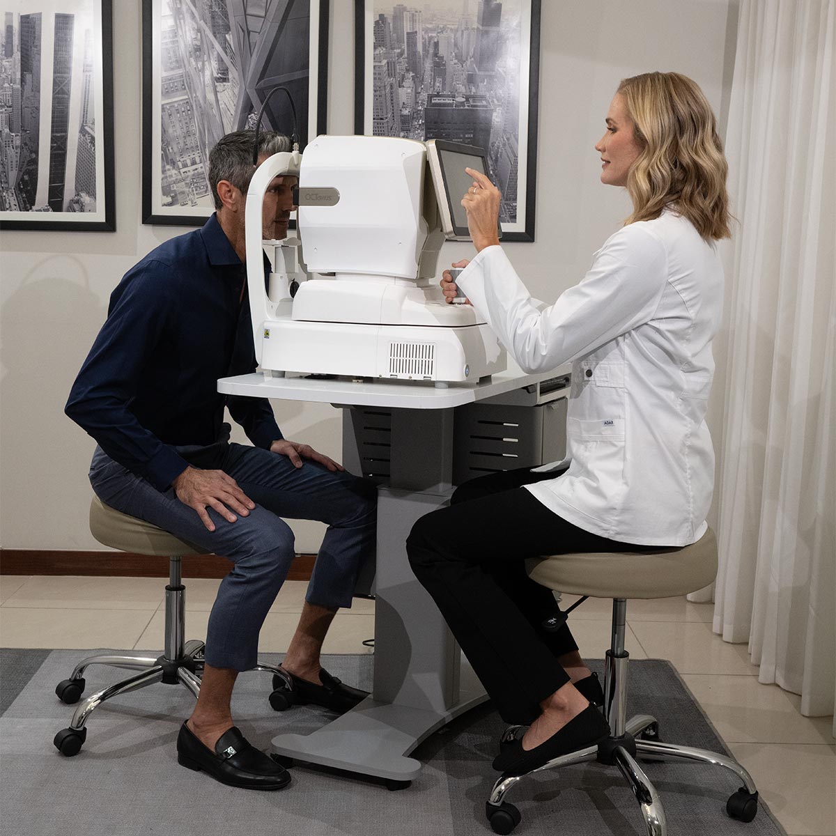
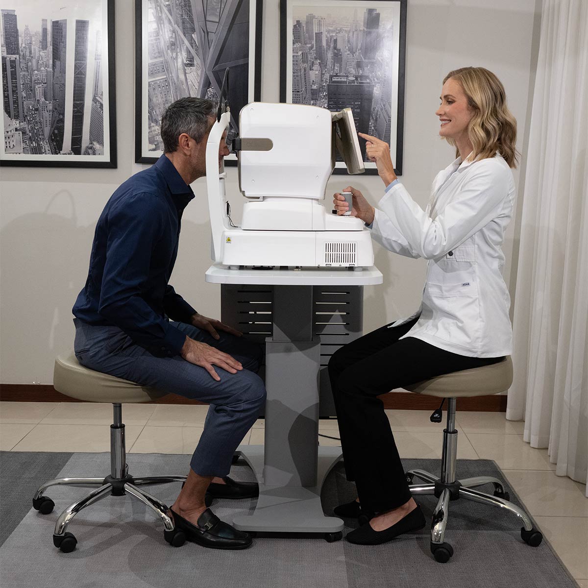
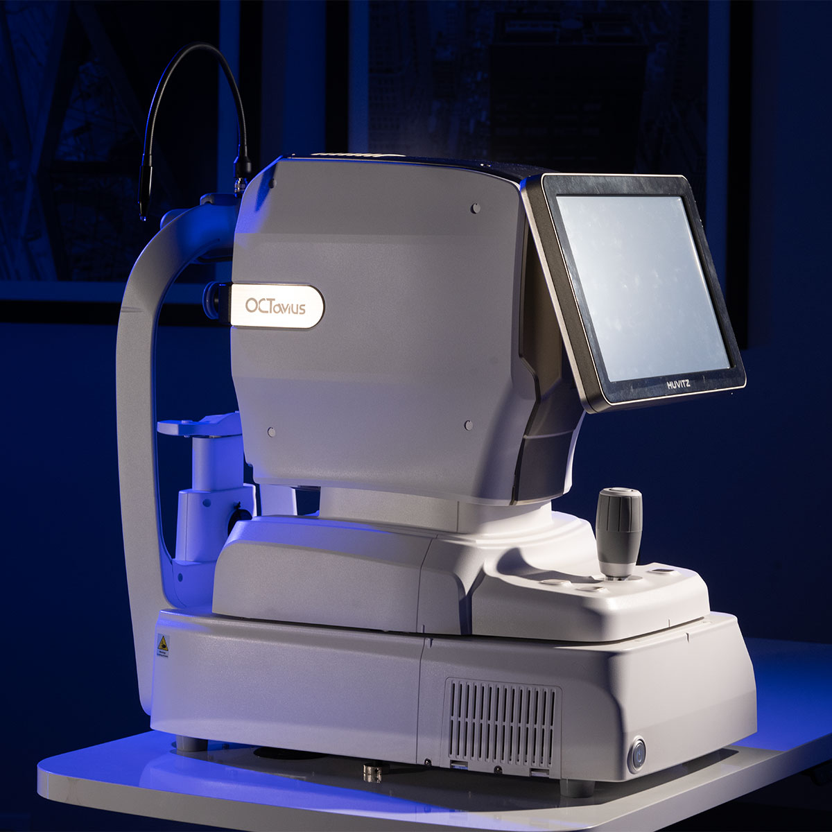
The HOCT-1F (80) is Huvitz’s next-generation OCT, designed to enhance scanning speed, angiography quality, and overall imaging performance. It now achieves 80,000 A-scans/sec (+17%), reducing patient waiting time and the need for repeat scans.

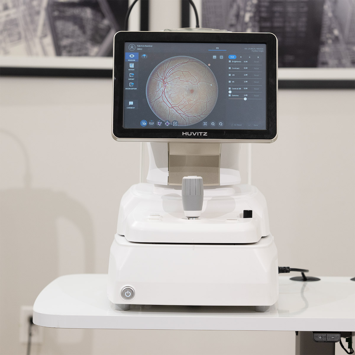
The Fundus Enhancement Lv.4 function, with refined brightness and gamma control, enables the visualization of subtle lesions, while Triple Angiography — featuring motion correction, retinal tracking, and noise reduction — produces sharper and more reliable images.

In addition, an enhanced algorithm for the anterior segment optimizes edge definition, ensuring greater measurement accuracy, and the built-in PC (13th-generation Intel + SSD) guarantees fast and stable workflows.

Talk to a specialist

3D OCT

Sharp details for precise retinal diagnostics.
Fundus Camera

Clear and comprehensive retinal imaging for clinical evaluation.
Angiography

Detailed visualization of retinal circulation.

Topography

Quantitative analysis for follow-up and decision-making.

Biometry

Reliable measurements for safe surgical planning.


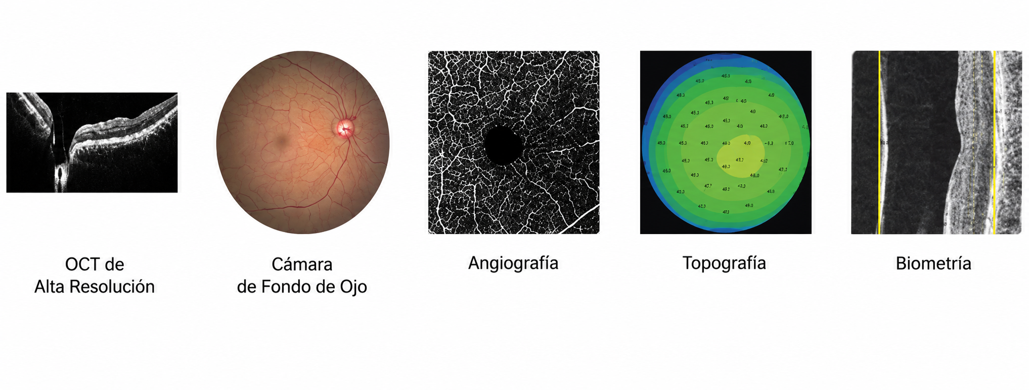
5-in-1 System: 3D OCT • Fundus Camera • Angiography • Biometry • Topography

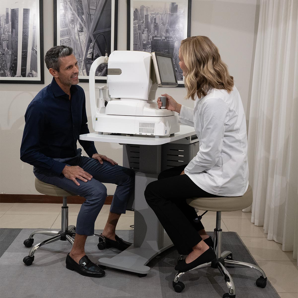
The HOCT-1F (80) integrates five essential diagnostics into a single platform, offering smooth operation and precise data that specialists can rely on. With this 5-in-1 system, you can increase diagnostic confidence, optimize workflow, and save valuable space in the examination room.


Compact Design with Integrated Intel® Technology

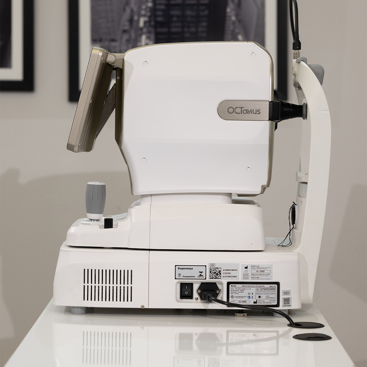
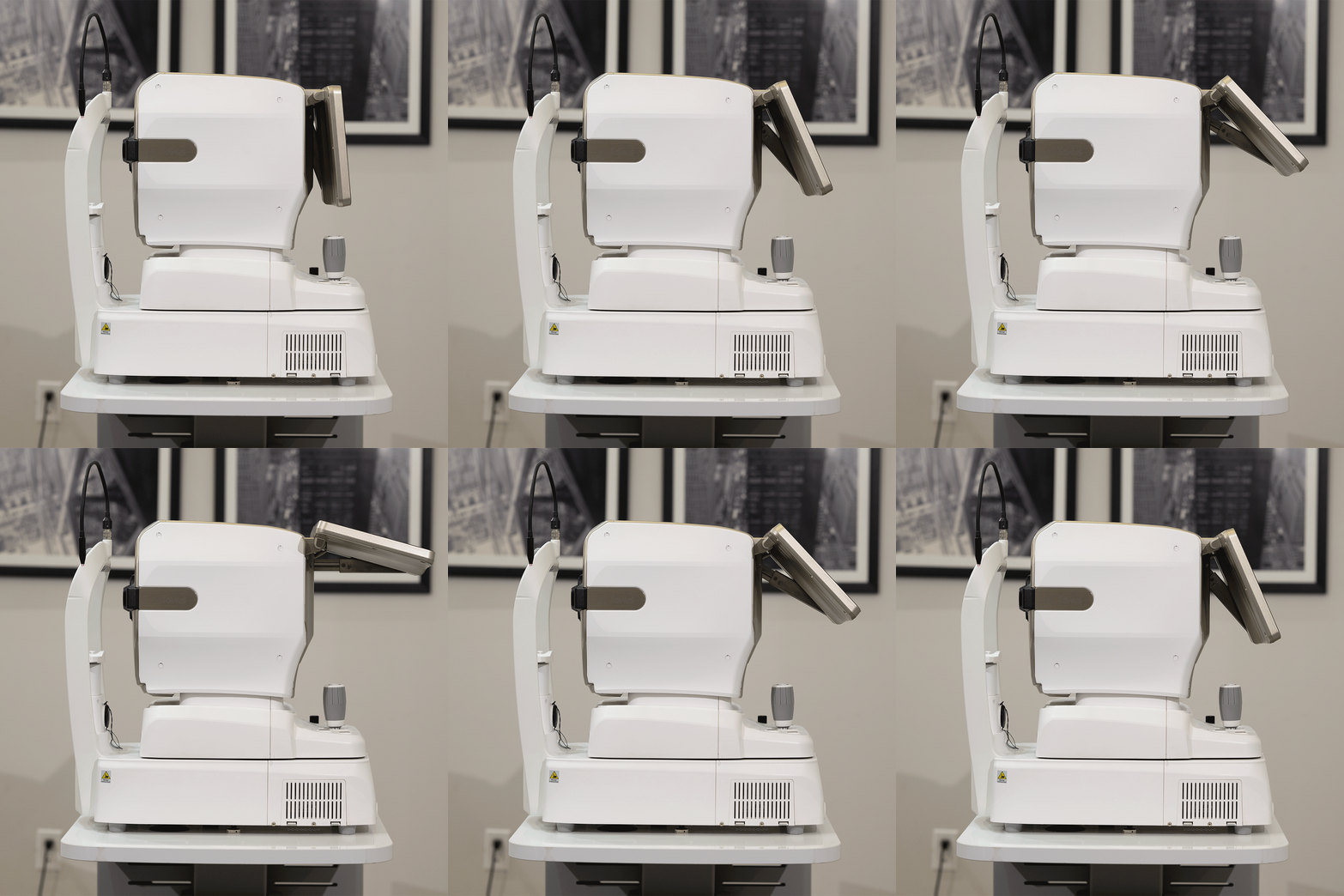
The HOCT-1F (80) incorporates an integrated computer powered by a 13th-generation Intel® i3 processor and a high-speed SSD unit, eliminating the need for an external PC and reducing installation footprint. Its true small footprint design makes it an ideal solution for modern clinics.

This integrated system delivers 35% faster processing, twice the speed for image startup and saving, along with enhanced reliability. The SSD ensures superior data durability and stability, while the integrated architecture provides a more organized and efficient workspace, with smooth performance for optimized clinical operations.

Key Features

The HOCT-1F (80) delivers superior performance with an enhanced scanning speed of 80K A-scans —a 17% increase over previous models— reducing patient wait times and optimizing workflow efficiency. Its 5-in-1 multifunctional design combines OCT, fundus imaging, angiography, topography, and biometry in a single compact system, streamlining diagnosis across multiple imaging modalities. Equipped with triple angiography, the HOCT-1F provides high-resolution visualization of the superficial, deep, and outer retinal layers, with advanced motion correction that eliminates the need for repeat scans. The Fundus Enhancement Lv.4 engine integrates Corneal Flare Removal, white balance, Domain Transform, central brightness, and gamma control to achieve exceptional image clarity and precise visualization of subtle lesions. An enhanced anterior B-scan increases accuracy in measuring the boundaries of the cornea and anterior chamber, reducing segmentation errors for more reliable thickness and radial maps. Finally, the built-in PC, powered by a 7th-generation Intel i7 processor and SSD storage, ensures faster boot times, agile image processing, and smoother overall performance, delivering an uninterrupted clinical experience.

High-Speed A-Scan Acquisition

With the A-scan speed increased from 68,000 to 80,000 A-scans/sec (+17%), the HOCT-1F (80) reduces patient waiting time and minimizes the need for repeated examinations by capturing stabilized, high-resolution 3D volumes even in the presence of micro-movements and blinking. Its advanced 80K engine delivers sharp, artifact-free images, facilitating operation even for new users and enabling detailed visualization of retinal layers for more confident diagnostic evaluations, including layer thickness analysis and macular differentiation.

High-Resolution Retinal and Choroidal Imaging

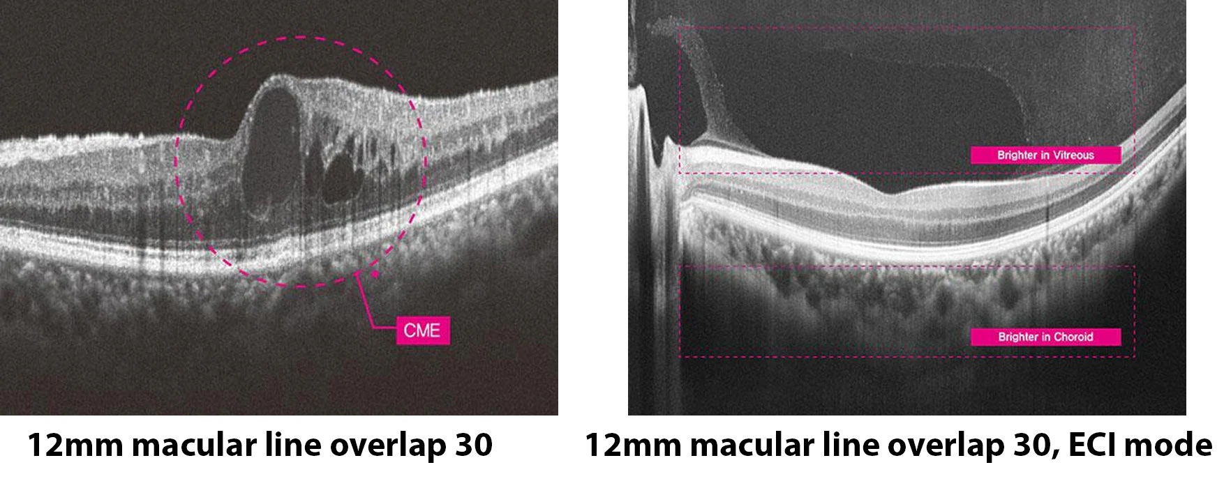
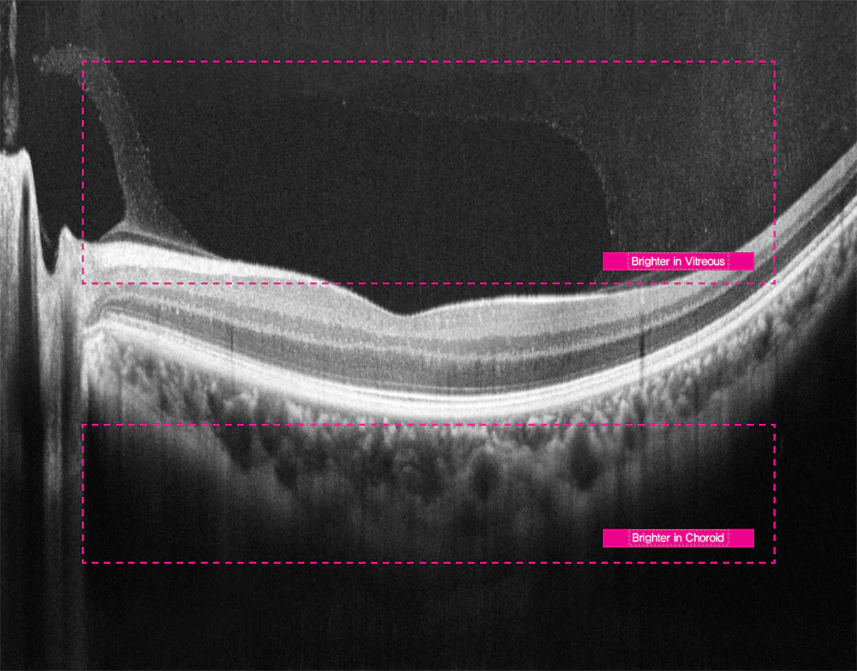
Capture the retina and choroid with unparalleled precision through high-resolution scans. The HOCT-1F (80) incorporates fast and stable scanning technology that delivers clear visualization of the delicate retinal and choroidal structures, proving highly effective in the diagnosis of critical ophthalmic conditions, such as retinal detachment and vitreous opacities.

In ECI (Enhanced Choroidal Imaging) mode, depth signals are inverted to maximize contrast within the choroidal layer, ensuring superior diagnostic clarity.

Image: ECI mode, 12 mm macular line with an overlap of 30.

Precise Capture of Microstructural Alterations

With exceptional clarity, the system detects subtle morphological anomalies, such as cystoid macular edema (CME), dome-shaped elevations, and early detachments of the RPE layer. This precision enables early and accurate evaluation of lesion structure and progression, supporting timely diagnosis and effective treatment planning.

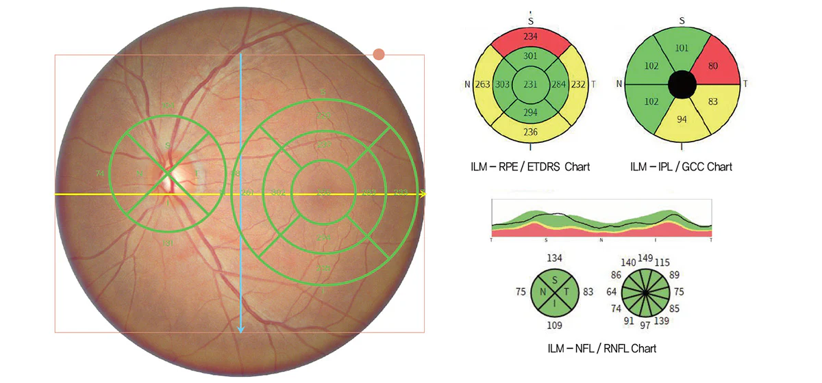
Simultaneous retina and optic nerve scanning

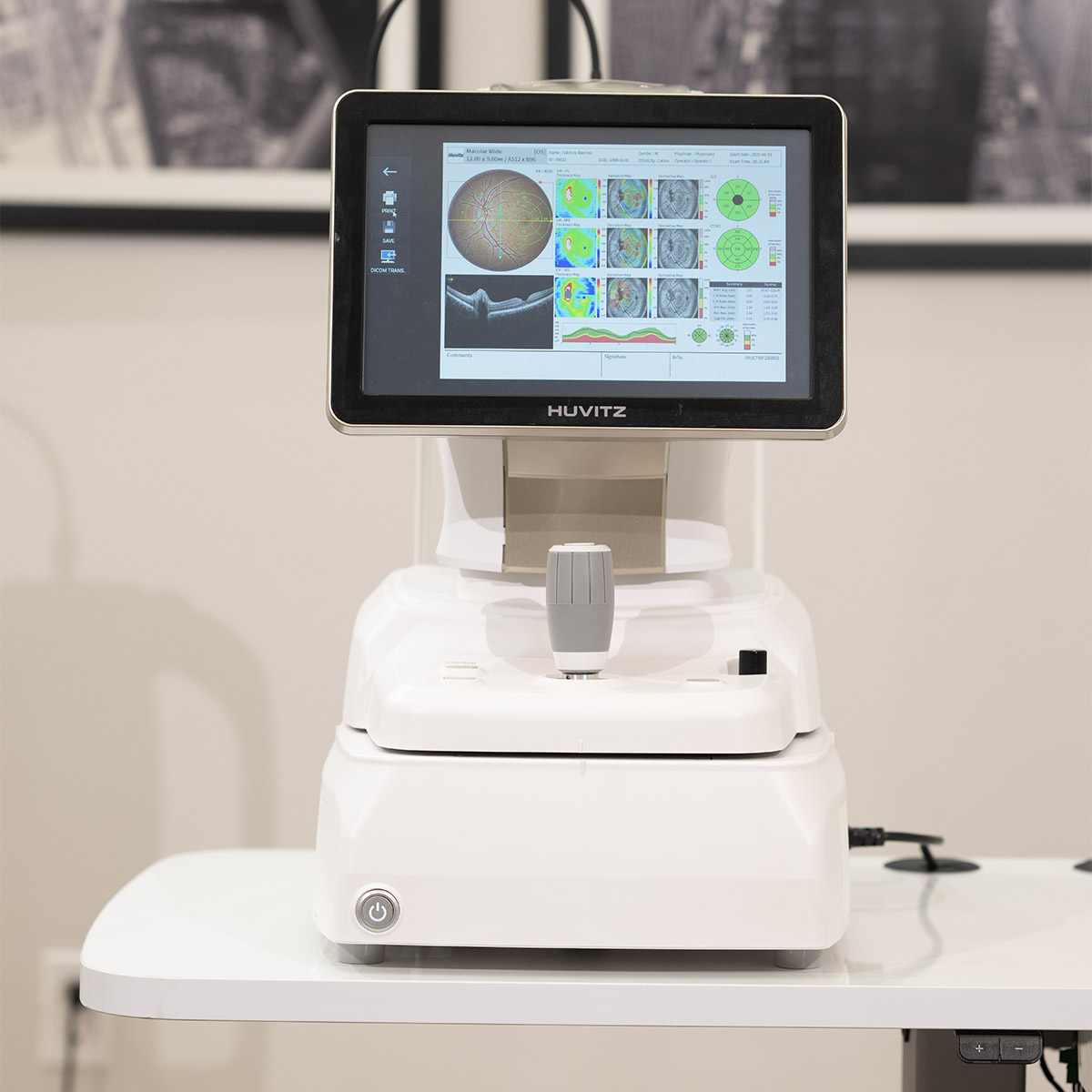
With a single 3D scan, the system captures both the retina and the optic nerve head, providing comprehensive analysis metrics such as the ETDRS chart, ganglion cell chart, RNFL chart, and TSNIT chart.

This integrated approach enables precise evaluation of structural alterations and facilitates early diagnosis of optic nerve-related conditions, including glaucoma, improving both diagnostic accuracy and efficiency.

Image: Wide macular line of 12 mm, overlap 10.

True Color Fundus in a Single Capture

Single Fundus Macula Level 4

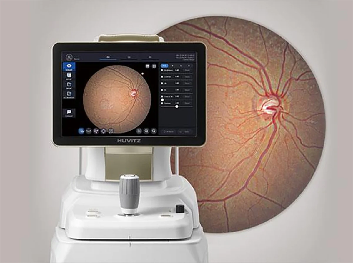
With 12-bit color depth and intelligent gamma correction, the HOCT-1F (80) captures true-color fundus images in a single exposure, with minimal chromatic distortion.

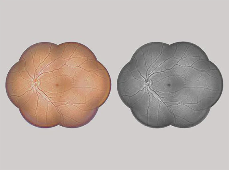
The system balances contrast between dark retinal areas and the bright optic disc, clearly delineating arteries, veins, and fine microvasculature. For broader coverage, it automatically aligns and combines up to seven images into a wide-field panorama, enabling intuitive identification of the location and extent of lesions in an integrated view.


Wide-angle panoramic vision for lesion monitoring

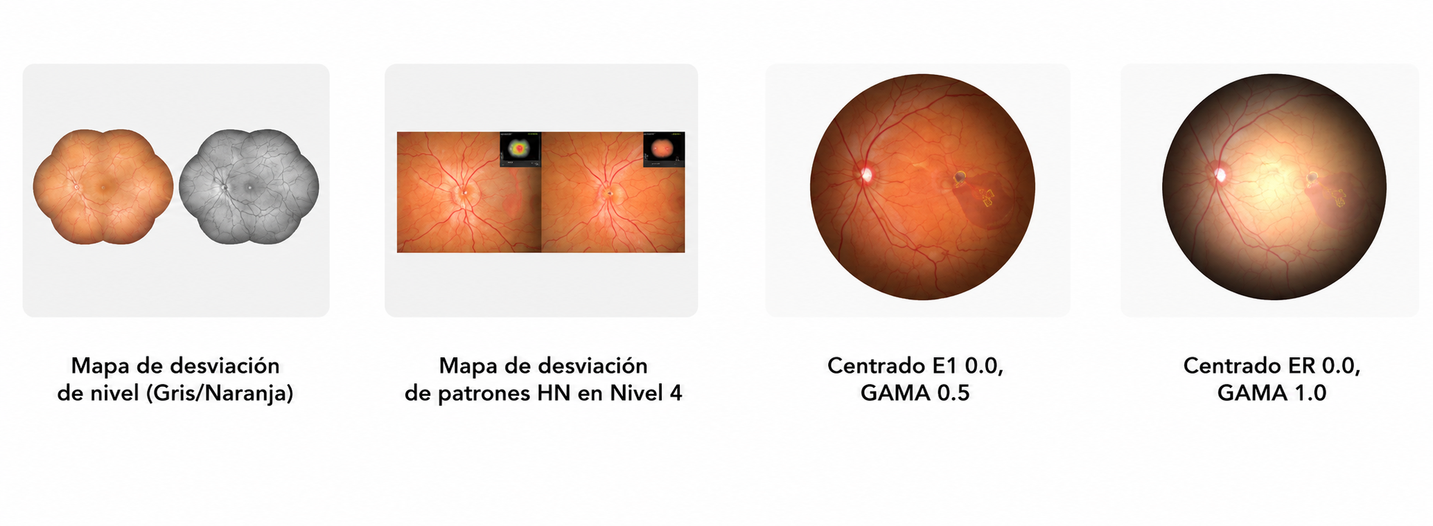
Generates a wide-angle panorama by combining 2 to 7 fundus images into a single display. This expanded field allows a clear visualization of the entire fundus structure, facilitating intuitive identification and precise monitoring of the location and extent of lesions, especially in conditions such as diabetic retinopathy.

Image: Fundus panorama, level 4 / grayscale.

Structural Analysis of the Optic Nerve Head

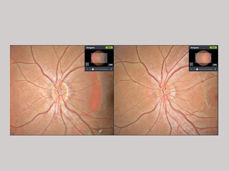
The system performs precise measurements of the optic nerve head from multiple left and right viewpoints. This multi-angle approach enables a highly accurate assessment of structural alterations and potential damage to the optic nerve head, providing reliable data for the early detection and monitoring of optic nerve-related diseases.

Image: Disc stereo (fundus) in HIIS-1, level 4.

Artificial intelligence analysis of 12 types of lesions

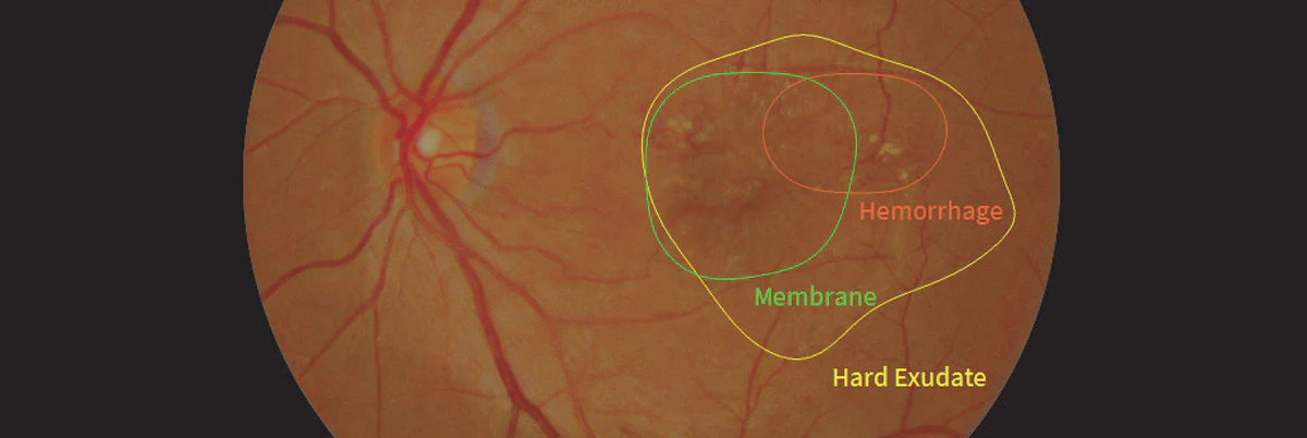
The advanced AI-based fundus analyzer divides the retina into eight distinct regions and evaluates 12 types of lesions with high precision. It accurately identifies and marks their locations, providing reliable data that supports faster, more accurate diagnoses and better-informed clinical decisions.

Image: Single macular fundus, level 1, VUNO AI image.

Fundus Image Enhancer Lv.4

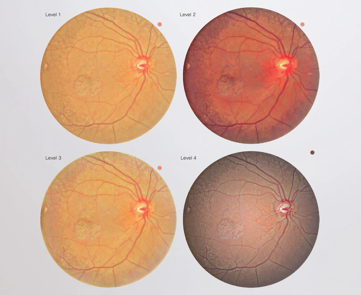
Offers five customizable enhancement modes —from the original image (Non) to levels 1, 2, 3, and 4— adapting to different clinical scenarios and user preferences.

By progressively adjusting central brightness and gamma, the system balances dark retinal areas with the bright optic disc, improves edge contrast, and highlights the microvasculature without altering true colors.

The result is sharper, more uniform fundus images that facilitate the detection of subtle findings, improve comparisons between examinations, and provide clear images for documentation and patient education.

At level 4, the maximum enhancement level, fine vascular details and retinal patterns are rendered with the highest clarity for more confident diagnostic interpretation.

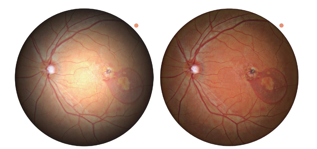
Enhanced lesion visibility through brightness and color adjustment

Al ajustar los valores de BR central y Gamma, es posible optimizar el brillo y el equilibrio de color de la imagen de fondo de ojo. Esta optimización permite una visualización más clara de áreas específicas, haciendo que las lesiones sean más definidas y fáciles de evaluar para un diagnóstico preciso.

Imagen: Agujero macular complicado con DMAE. Fondo de ojo macular único, nivel 4, Universidad Dankook (con opciones actualizadas de brillo central aplicadas).


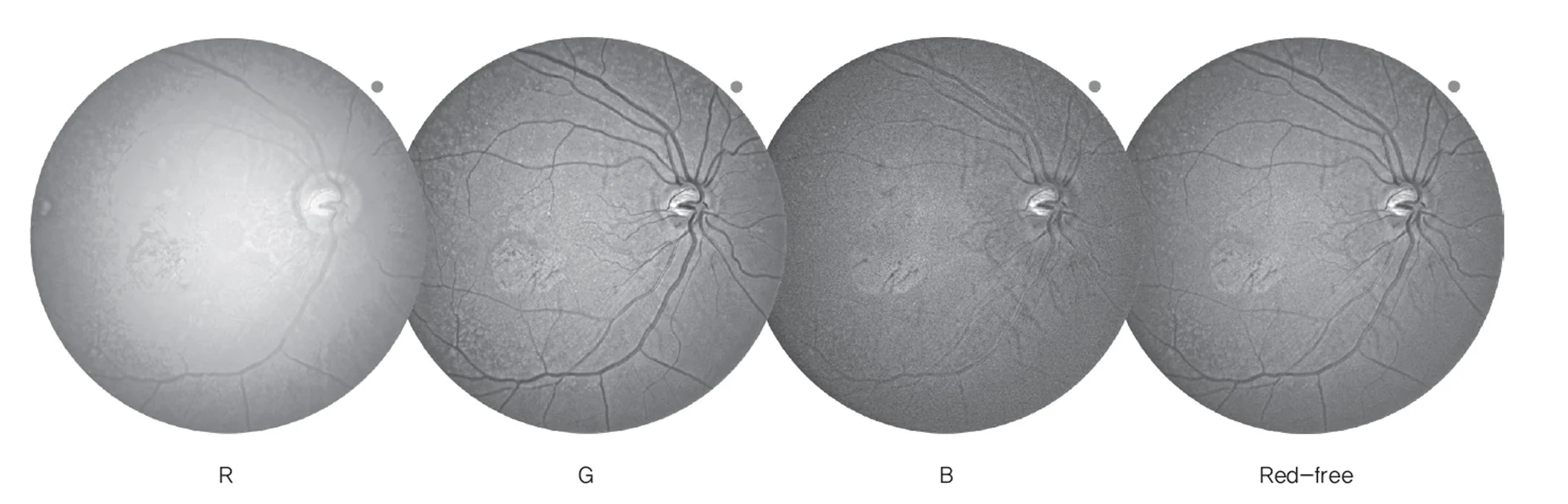
Unique macular fundus image, level 4, Dankook University (R/G/B channel mode).

Customizable RGB Color Channels

Users can enhance visualization by selecting the R (Red), G (Green), or B (Blue) color channels, allowing targeted observation of lesions in the most suitable color spectrum for a more precise evaluation.

Clinical Insight

R Channel:
used to observe structures reaching the deep retinal layers and the choroid (analyzes deep retinal vessels, choroidal abnormalities, and hemorrhagic lesions).

G Channel:
used to express the contrast of superficial vessels and the nerve fiber layer (diagnoses vascular changes such as retinal edema, microhemorrhages, and neovascularization).

B Channel:
used to visualize fine structures of the retinal surface and the nerve fiber layer (aids in the early diagnosis of nerve fiber layer damage, such as glaucoma).

Enhanced Angiography — Fast OCT-A Imaging, Full Coverage

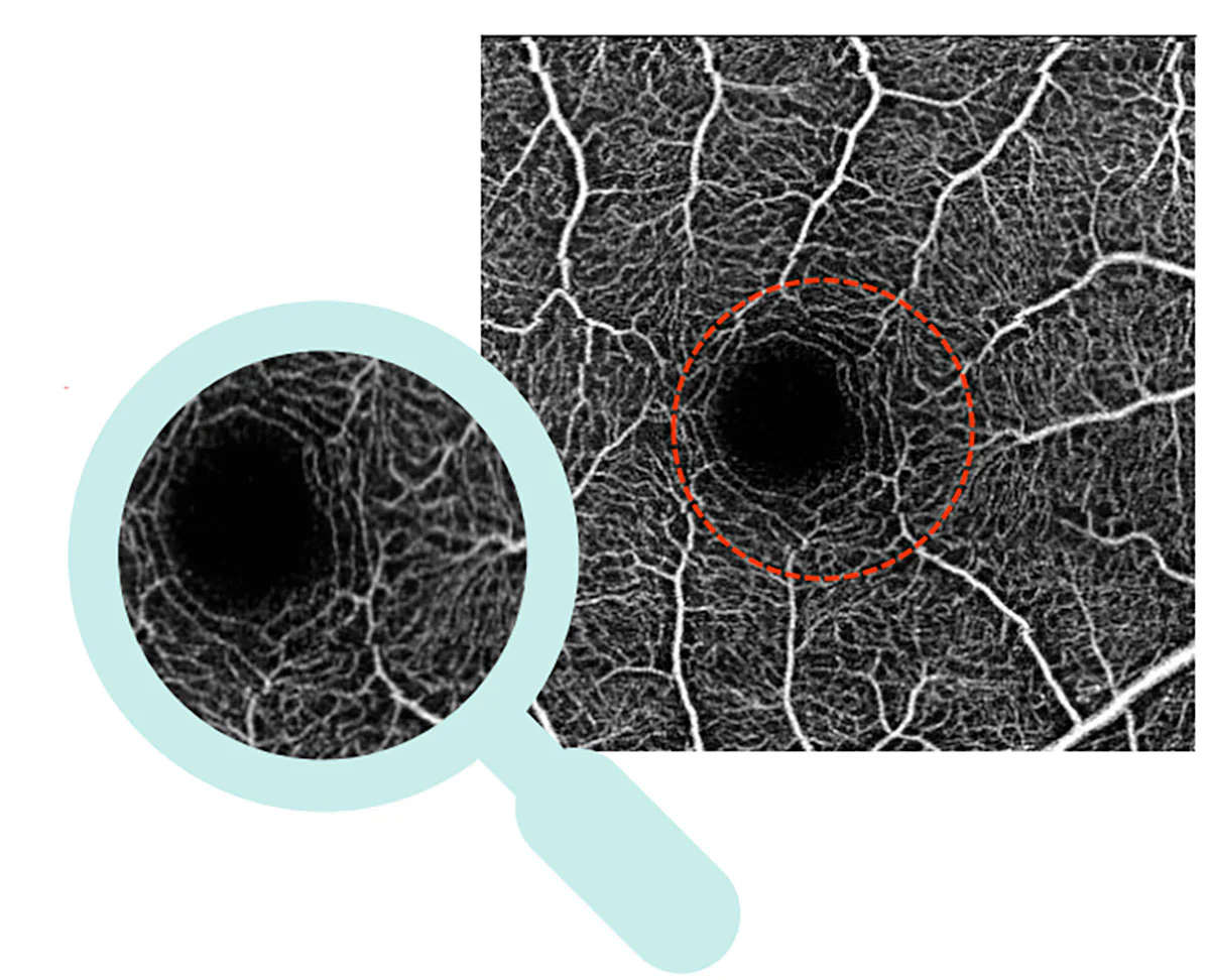
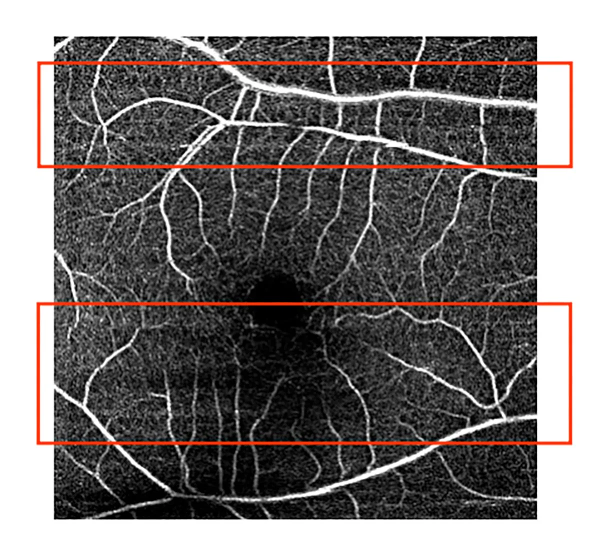
The Angio Enhancement suite of the HOCT-1F (80) increases performance and reliability by combining scan speed increments based on location with real-time retinal tracking via a 30 fps infrared (IR) camera. Image quality is elevated through slab-based vascular signal contrast with foveal reflex suppression, along with post-acquisition B-scan registration that restores interrupted vessels. CNV (choroidal neovascularization) visualization benefits from an improved despeckling process that highlights only true neovascular structures. Robust noise controls eliminate motion artifacts, suppress horizontal banding noise, and remove the ghosting effect, while PAR (projection artifact removal) eliminates superficial vessel shadows and more sharply defines the outer retina. Finally, refined retinal layer segmentation enables more precise analysis of regions in OCTA, reinforcing diagnostic confidence.

Real-Time Retinal Tracking

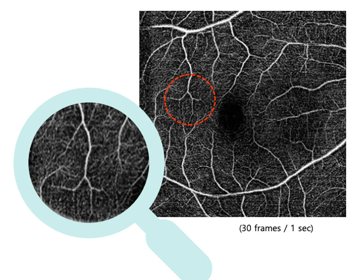
The HOCT-1F (80) offers precise visualization of the retinal microvasculature through real-time retinal tracking with a 30 fps IR camera that corrects eye movement during acquisition.

By reducing motion blur and suppressing banding artifacts, it preserves vascular continuity and sharp capillary borders, producing stable and faithful OCTA images even in the presence of small fixation movements or blinks, allowing clearer data to be captured with fewer repetitions.

Image: 30 frames / sec

Enhanced Signal Contrast in CNV

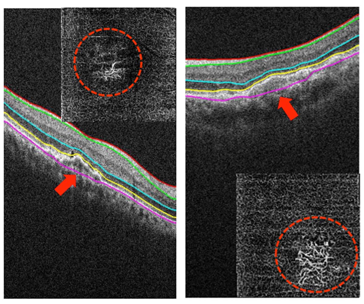
The HOCT-1F (80) improves the visibility of choroidal neovascularization (CNV) in the outer retina by optimizing signal processing within the selected OCTA slab, suppressing foveal specular reflections and reducing noise.

The result is greater vessel-to-background contrast with preserved microvascular continuity, a cleaner representation of fine capillaries, and more reliable delineation of CNV networks, enhancing detection, boundary assessment, and comparison in clinical follow-up.

Motion Artifact Removal

The HOCT-1F (80) minimizes motion-related artifacts in OCTA by correcting ocular micro-movements during acquisition and applying a dedicated “stripe noise” reduction algorithm that suppresses horizontal banding.

The result is cleaner slabs with preserved vascular continuity, sharper capillary edges, and reduced background noise, providing more readable images and reducing the need for repeated scans.

Projection Artifact Removal (PAR)

The HOCT-1F (80) uses PAR to suppress shadow-like projections from the superficial vascular plexus that contaminate deeper OCTA slabs.

By eliminating these projection artifacts, the system preserves genuine flow signals in the outer retina, improves vessel-to-background contrast, and maintains microvascular continuity.

The result is a cleaner, more faithful visualization of structures such as CNV, with sharper borders and fewer false positives, supporting more accurate interpretation and more reliable longitudinal quantification.

Enhanced Retinal Layer Segmentation (RPEDC)

The HOCT-1F (80) delivers more precise layer segmentation, with special improvements in the RPE/Bruch’s membrane complex. Its refined algorithm reliably tracks the RPEDC boundary, even in irregular morphologies, reducing manual corrections and increasing reproducibility.

By clearly separating the outer retinal layers, it enables better visualization of sub-RPE structures, improved detection and delineation of drusen/PED, and more reliable thickness and en-face maps.

The result is consistent slab selection and greater diagnostic confidence in the evaluation of subtle RPEDC changes over time.

Topography

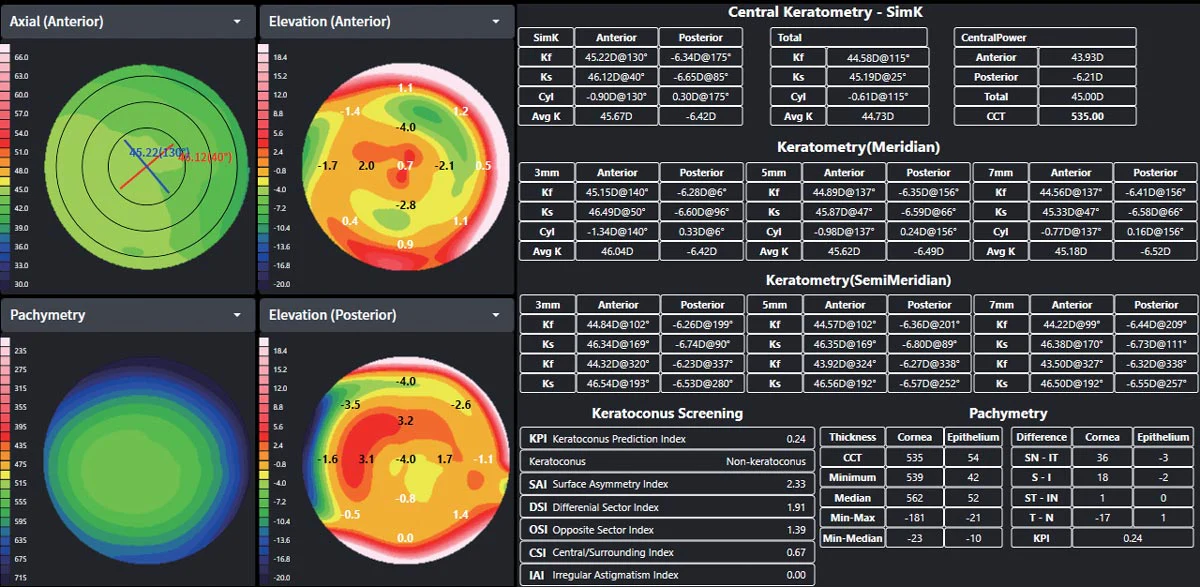
The OCT topography simultaneously captures the anterior and posterior corneal surfaces, generating a detailed 3D analysis with 16 types of corneal maps for precise evaluation of thickness, curvature, and elevation.

Maps and numerical data on a single screen

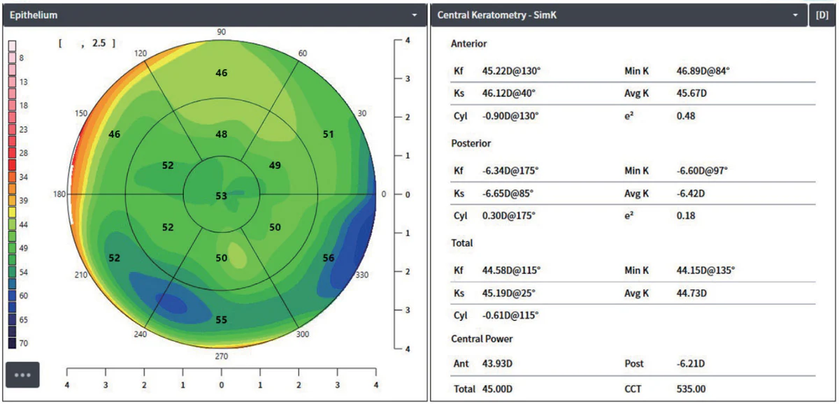
On a single screen, the user can visualize four maps — Axial, Anterior Elevation, Posterior Elevation, and Pachymetry — while analyzing key parameters such as SimK, Meridian, Keratoconus Index, and epithelial thickness.

Maps can be changed or the layout adjusted as needed, allowing flexible use tailored to the diagnostic purpose and situation.

Simplified clinical workflow

Analysis screens can be printed or sent to a PACS server in DICOM format, facilitating the storage and exchange of results. When printing, the background can be changed to white to improve report readability. Integration with EMR and hospital PACS systems further streamlines the clinical workflow.

Biometry

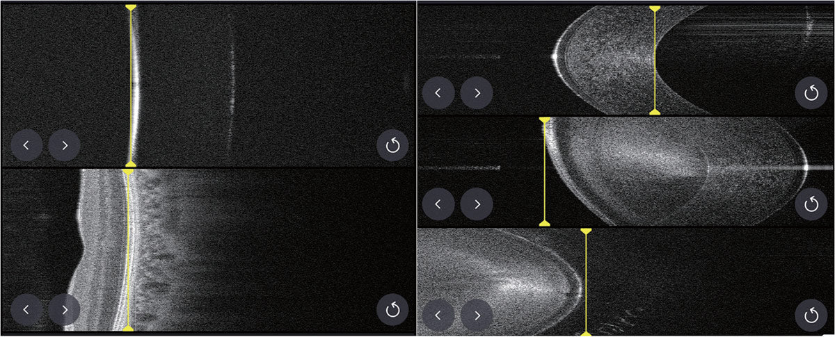
As a complement to topography, optical biometry visualizes the entire axial length —from the cornea to the macula— in high-resolution 2D, providing richer detail than conventional ultrasound. Together, they deliver the precise data needed for accurate, personalized IOL selection for each patient and for surgical planning with complete confidence.

Fast, accurate, and highly reliable measurements

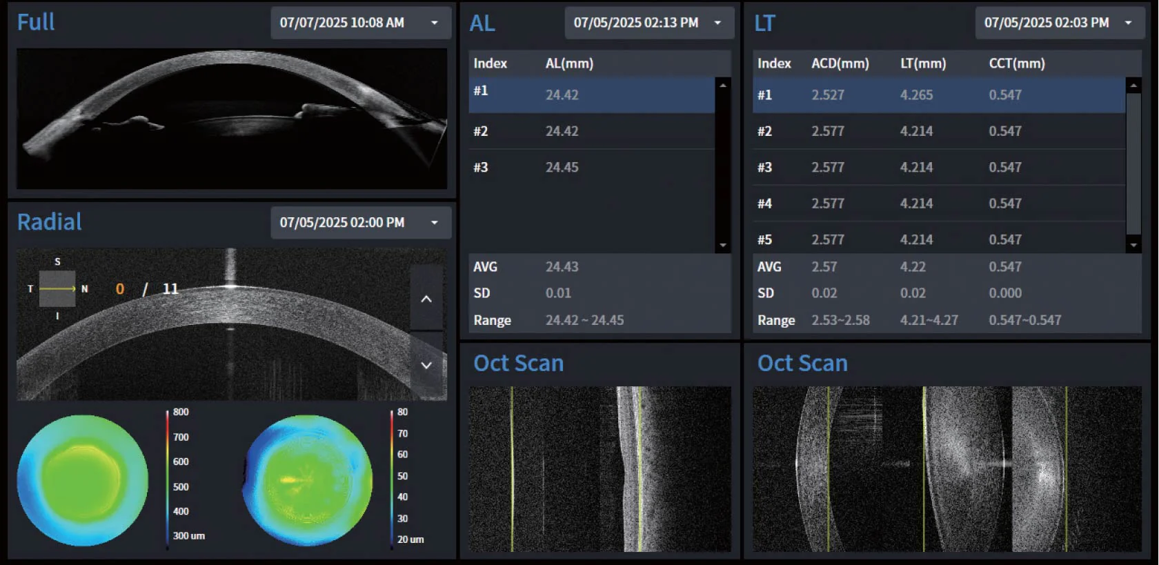
With Burst Mode, the system performs rapid consecutive measurements —three times for axial length and five times for each of the following parameters: central corneal thickness (CCT), anterior chamber depth (ACD), and lens thickness (LT)— ensuring superior consistency and accuracy.

After each scan, B-scan images can be reviewed immediately on the confirmation screen, allowing fine adjustments when necessary. Low-quality scans caused by blinks or small eye movements are automatically filtered out, ensuring reliable IOL calculations and precise surgical planning, even in challenging cases involving dense cataracts or macular distortion.

Complete Vision: From Precise Measurements to IOL Recommendations

The biometry analysis screen provides a comprehensive three-dimensional view of ocular structures with AL, TL, Radial, and Full Anterior Segment perspectives.

Measurement parameters such as axial length (AL) and lens thickness (TL) can be selectively reviewed, with the option to manually adjust layer boundary markers for maximum precision, even in complex anatomies.

By integrating AL, CCT, ACD, LT, and K values based on corneal topography, the system automatically generates ideal IOL recommendations tailored to each patient, supporting precise and efficient surgical planning.

Enhanced Anterior Imaging

OCT Topography simultaneously captures the anterior and posterior surfaces of the cornea, producing a detailed 3D analysis with 16 types of corneal maps for precise evaluation of thickness, curvature, and elevation.

Complementing this technology, Optical Biometry visualizes the entire axial length — from the cornea to the macula — in high-resolution 2D images, offering significantly more detail than conventional ultrasound.

Together, these functionalities provide detailed and accurate data for personalized IOL selection and safer, more reliable surgical planning.

Complete Anterior Imaging for Comprehensive Visualization

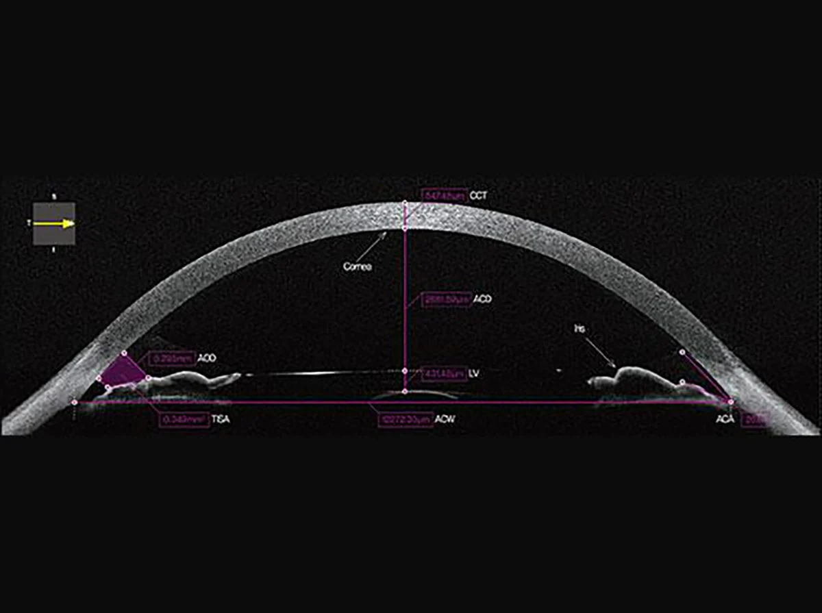
A single scan captures the entire anterior segment —from the cornea to the lens— with clear visualization of the iris and anterior chamber. Essential parameters such as WTW (white-to-white) and ACT (anterior chamber thickness) are measured automatically, enabling objective comparisons and precise evaluations.

This comprehensive image supports accurate differentiation between open-angle and angle-closure glaucoma, as well as detailed pre- and postoperative assessments in refractive or lens surgery.

Clinical Insight: Useful for the early detection of anterior chamber angle abnormalities and for pre- and postoperative monitoring in procedures involving refractive lenses.

Ciliary Muscle and Lens Visualization

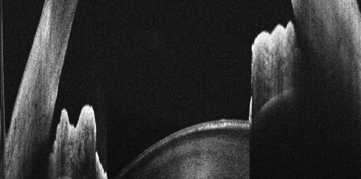
With the Wide One Line Scanning function, the anterior structure of the ciliary muscle and lens is clearly visualized in a single scan. This enables rapid assessment of alterations in ciliary muscle function, as well as the position and shape of the lens, making it ideal for pre- and postoperative comparisons.

It also supports detailed functional analysis of the anterior segment across a variety of clinical applications, including presbyopia correction and IOL implantation.

Clinical Insight: Rapid evaluation of ciliary muscle function and changes in lens position for pre- and postoperative comparisons.

Corneal Analysis

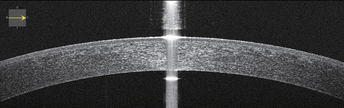
With the Anterior Segment Module, high-resolution cross-sectional images of the cornea are captured to precisely measure thickness, curvature, and anterior and posterior structures. These measurements can be visualized and analyzed in 2D and 3D formats, providing comprehensive data for accurate clinical evaluations.

Radial Analysis

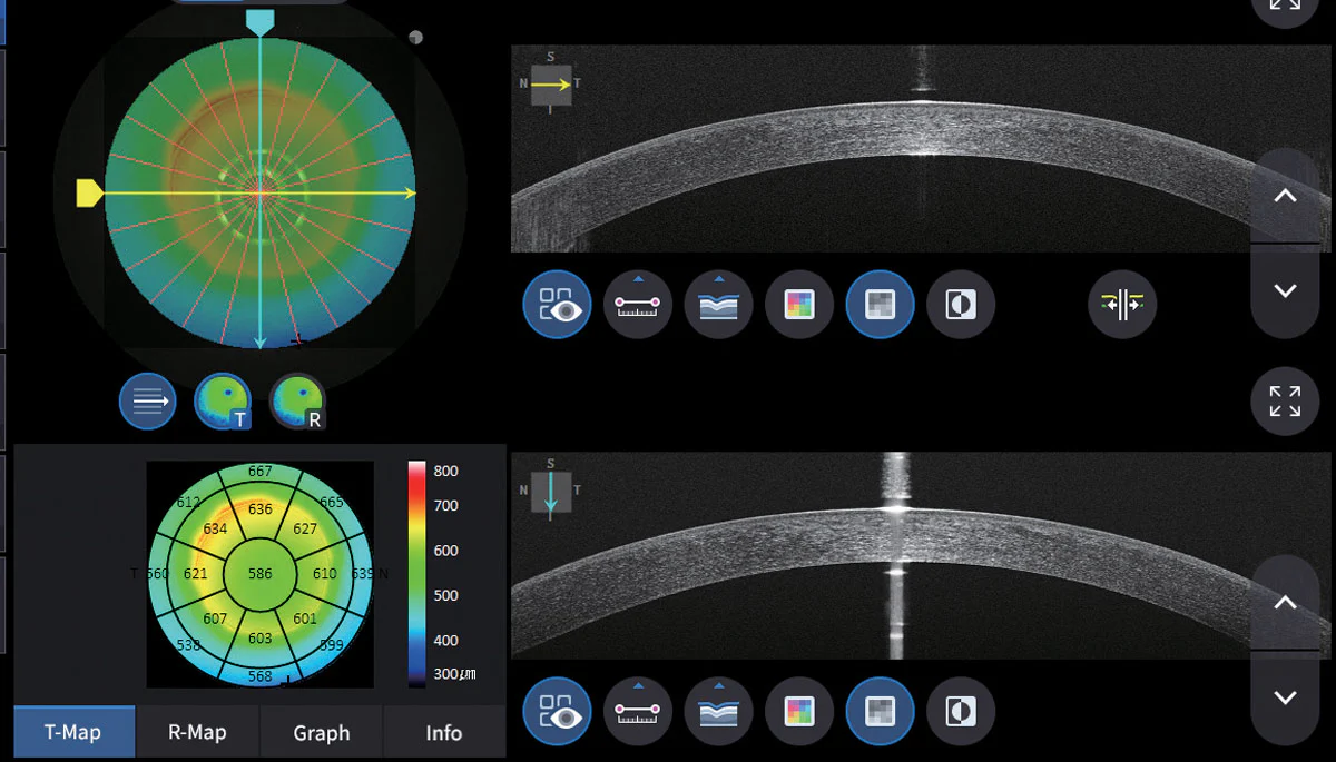
Utilizing the Corneal Thickness Map, the global thickness distribution and the location of the thinnest point are precisely identified.

This detailed visualization of corneal topography aids in the early detection and evaluation of anomalies such as keratoconus.

Anterior Chamber Angle Analysis

Mediante imágenes seccionales del segmento anterior, el sistema mide con precisión el ángulo de la cámara anterior (ACA) entre la córnea y el iris. Esto permite evaluar con exactitud si el ángulo está abierto o cerrado, respaldando un diagnóstico y seguimiento confiables de las condiciones relacionadas con el ángulo.

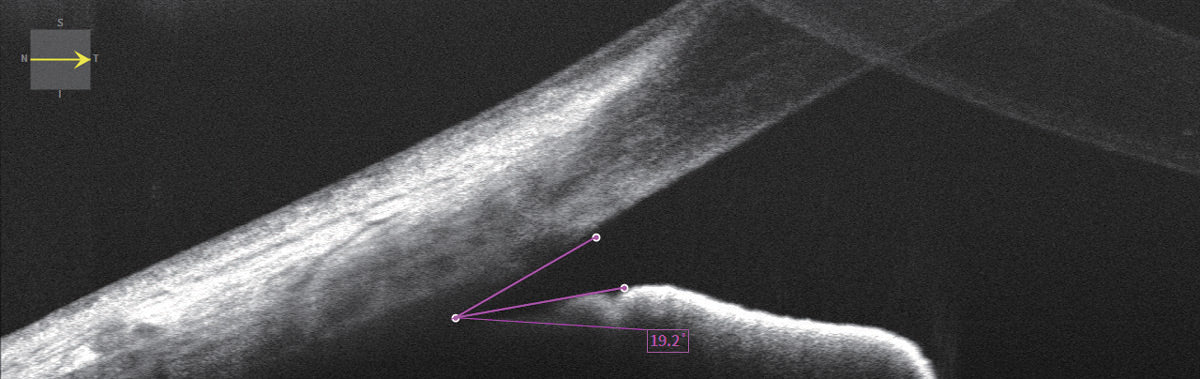
Anterior Chamber Angle Analysis

Through cross-sectional images of the anterior segment, the system precisely measures the anterior chamber angle (ACA) between the cornea and the iris. This enables accurate assessment of whether the angle is open or closed, supporting reliable diagnosis and monitoring of angle-related conditions.

More Reliable Maps – Parameters with Greater Precision

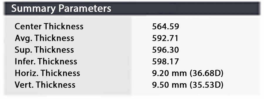
The HOCT-1F (80) generates more consistent and reliable maps, improving the accuracy of essential corneal parameters, including central thickness, average thickness, and curvature measurements.

These precise values are fundamental for advanced diagnostics and truly personalized patient care.


Specifications

Manuals, Catalogs & Clinical Research

Catalogs

Huvitz Optical Coherence Tomographer with Fundus

Manuals

Huvitz Optical Coherence Tomographer with Fundus

Clinical Research

Huvitz Optical Coherence Tomographer with Fundus