








Clinical Areas:

Retina and Fundus

Glaucoma and Intraocular Pressure

Myopia Control

Cataract

  • Home
  • Tomey Corneal and Anterior Segment Optical Coherence Tomography (OCT) CASIA2

Tomey Corneal and Anterior Segment Optical Coherence Tomography (OCT) CASIA2

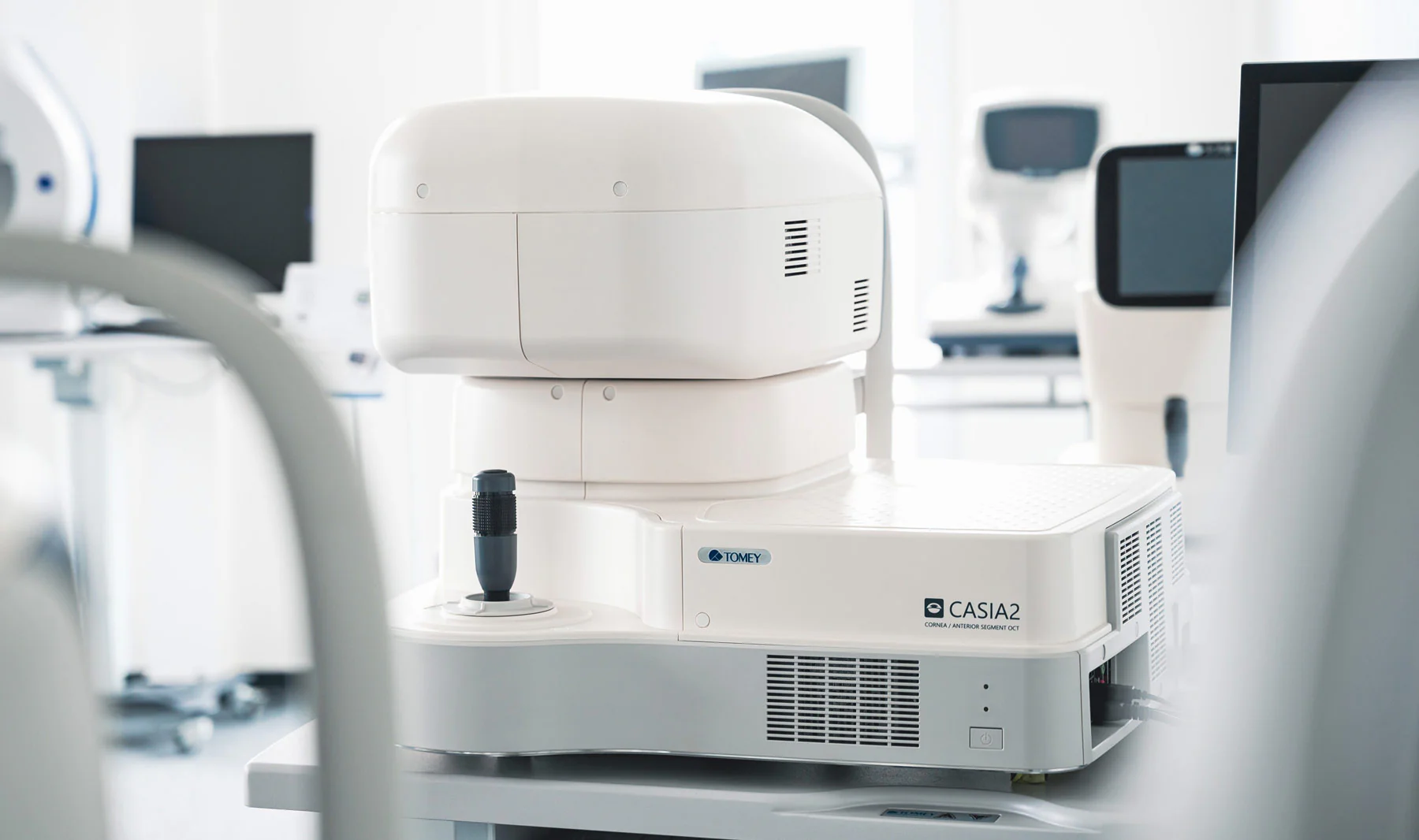
The **CASIA2** is the most advanced device for anterior segment analysis of the eye. The CASIA2 combines Placido disc corneal topography with a high-resolution OCT-based anterior segment tomography.

The **CASIA2** is the most remarkable product for all its distinct functions, capabilities, and outstanding technology.

The **CASIA2** is recognized for producing cross-sectional images with the highest resolution and clarity, with a diameter of 16 mm, along with numerous details of the corneal structure and layers. The CASIA2 can measure and detect on the Placido image, so that elevation, slope, and curvature data can be calculated using the arc-step method with conic curves. The anterior cornea, posterior cornea, anterior lens, and iris profiles are obtained from SD-OCT scans.
Talk to a specialist

Ultra-Fast Scanning

Captures 50,000 A-scans per second for detailed anterior segment imaging.

360° Glaucoma Analysis

Evaluates the complete angle with precision for a comprehensive glaucoma diagnosis.

Complete Cataract Management

Guides the pre- and postoperative process with advanced IOL selection tools.

Total Diagnostic Precision

Analyzes the crystalline lens, cornea, and anterior segment with color-coded parameters.

The Tomey CASIA2 is a high-precision anterior segment imaging diagnostic solution, designed for advanced applications in cataract, glaucoma, and corneal surgery. Featuring 360° angle analysis for glaucoma, high-speed scanning (50,000 A-scans/second), and a scan depth of up to 13 mm, the CASIA2 delivers detailed, high-resolution images. Key functions include corneal topography, phakic IOL simulation, crystalline lens shape analysis, intraocular lens selection calculations, and comprehensive tools for pre- and postoperative evaluations, including ICL. All of this makes it an all-in-one tool for thorough and accurate clinical assessment.

Cataract

Cataract Application

The Pre-OP Cataract application guides you through the ideal preparation for cataract surgery, displaying the essential detection parameters for IOL selection.

The Post-OP application clearly visualizes and documents surgical quality, providing deeper insight into outcomes. The new color camera enhances toric IOL alignment checks by displaying toric markers in various modes.

Additionally, a new scan type (IOLSCAN) has been added to the Post-OP Cataract application, offering improved IOL visualization.

Total Analysis

The images and parameters of the anterior segment of both eyes can be reviewed at a glance. In addition to conventional preoperative and postoperative examinations, the CASIA2 facilitates routine clinical operations such as initial examinations, follow-ups, and informed consent discussions.
Parameters are color-coded to highlight differences between the left and right eye, or when values fall outside standard ranges.

Lens Analysis

The exclusive Lens Analysis application provides you with all relevant lens parameters, including anterior and posterior radial curvature, as well as lens tilt and decentration. This tool ensures an accurate prediction of your surgical outcome.

Pre-OP ICL
With two integrated size recommendation formulas, this application helps confirm the appropriate ICL sizing for surgery.

Postoperative Application

The Postoperative application clearly visualizes and documents the quality of your cataract surgery with all the necessary information for its ideal review. GLAUCOMA

Glaucoma

STAR 360° and Glaucoma Application

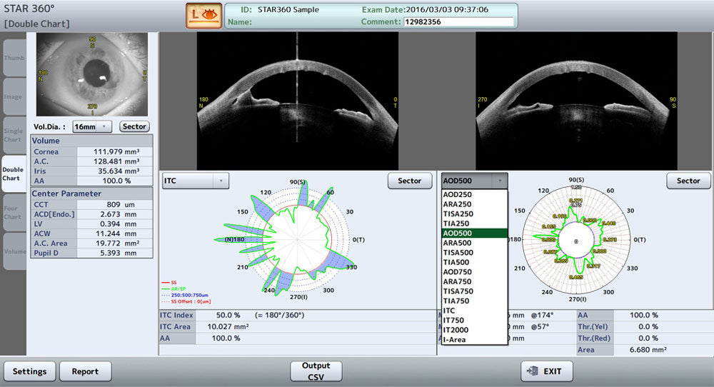
With our STAR 360° application, the CASIA2 automatically measures the anterior chamber angles throughout the entire anterior segment thanks to its automatic scleral spur detection, providing all the detailed parameters for an ideal glaucoma diagnosis.

Not only does it measure, but it also displays the results in a highly detailed and unparalleled chart. The charts can be selected according to your needs from all measured values.

All regions of interest, no matter how small or in which peripheral area they may be located, can also be scanned using a raster scan method, resulting in a fantastic scan density with high resolution.

Cornea

Corneal Application

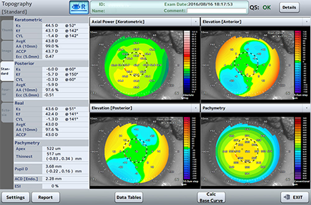
A topographic image is captured in just 0.3 seconds, ensuring the complete absence of artifacts. The cornea can be visualized in different types of maps with a wide range of individual configurations, either in 10 mm or 16 mm diameter. Scleral lens fitting is no longer an issue.

<Trend Analysis Application

Our exclusive Trend Analysis application visualizes the chronological progression of post-treatment regression of the eye, enabling highly precise monitoring. Different parameters can be selected for this chart.


Specifications

Manuals & Catalogs

Catalogs

Optical Coherence Tomography (OCT) CASIA2

Manuals

Optical Coherence Tomography (OCT) CASIA2