










Clinical Areas:

Retina and Fundus

Glaucoma and Intraocular Pressure

Myopia Control

Cataract

  • Home
  • Frey Perimeter AP-50

Frey Perimeter AP-50

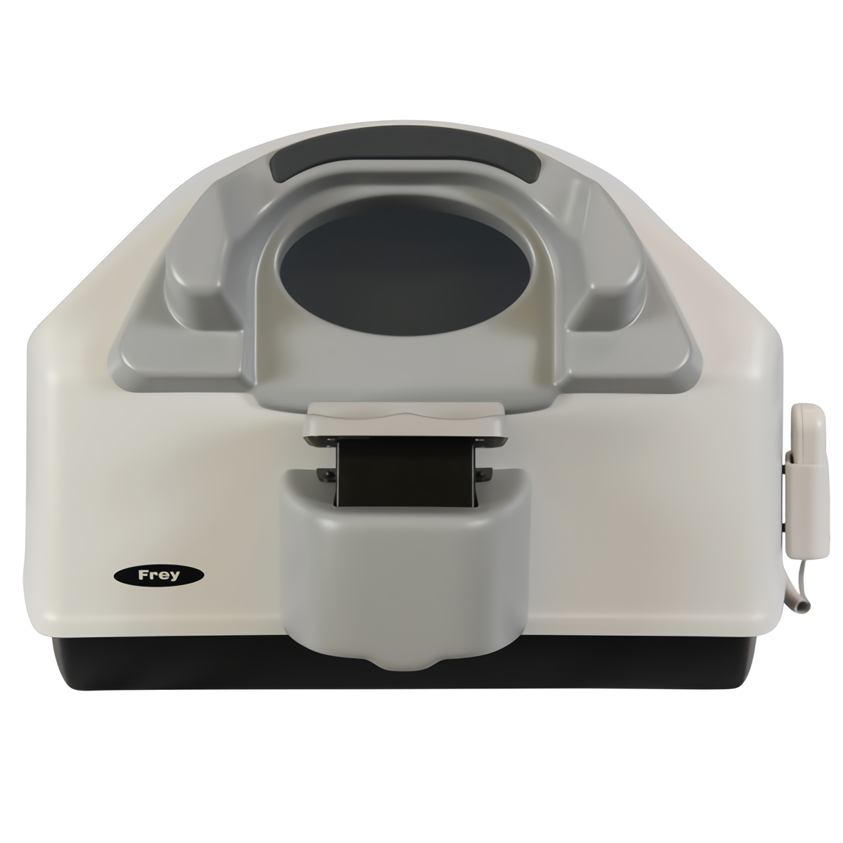
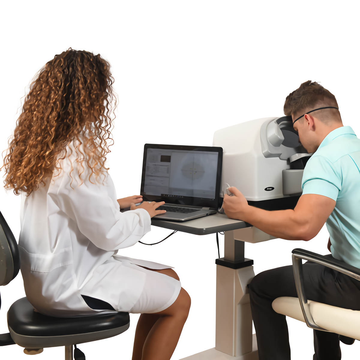
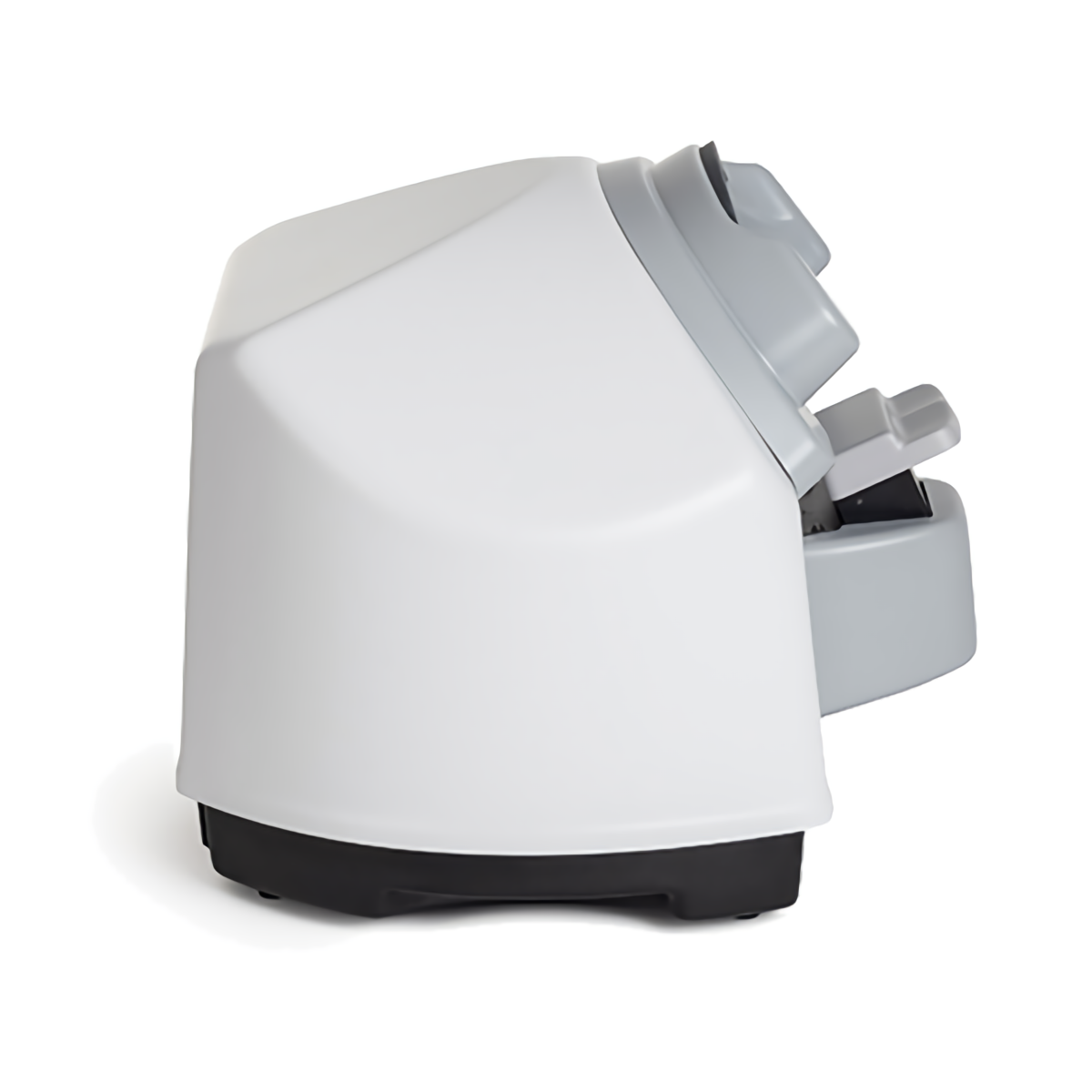
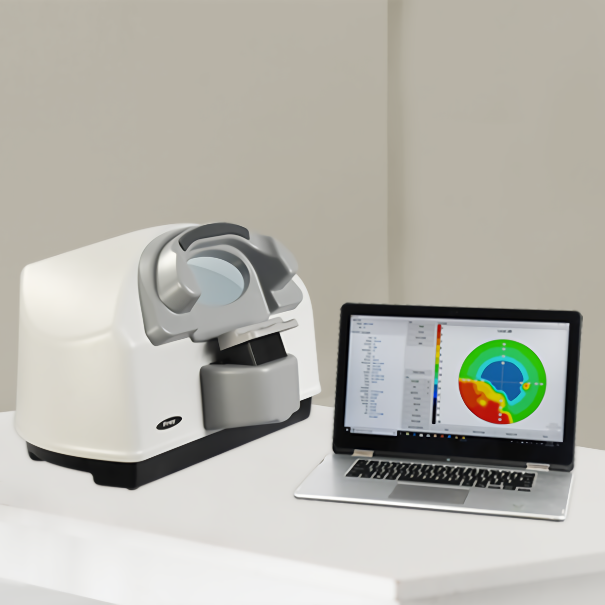
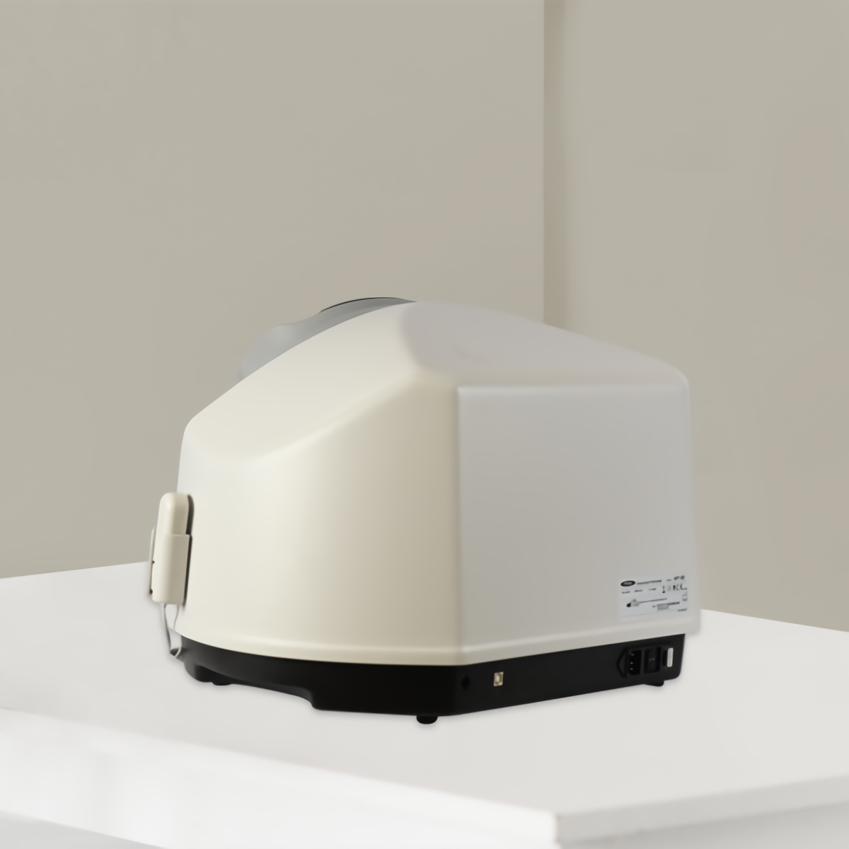
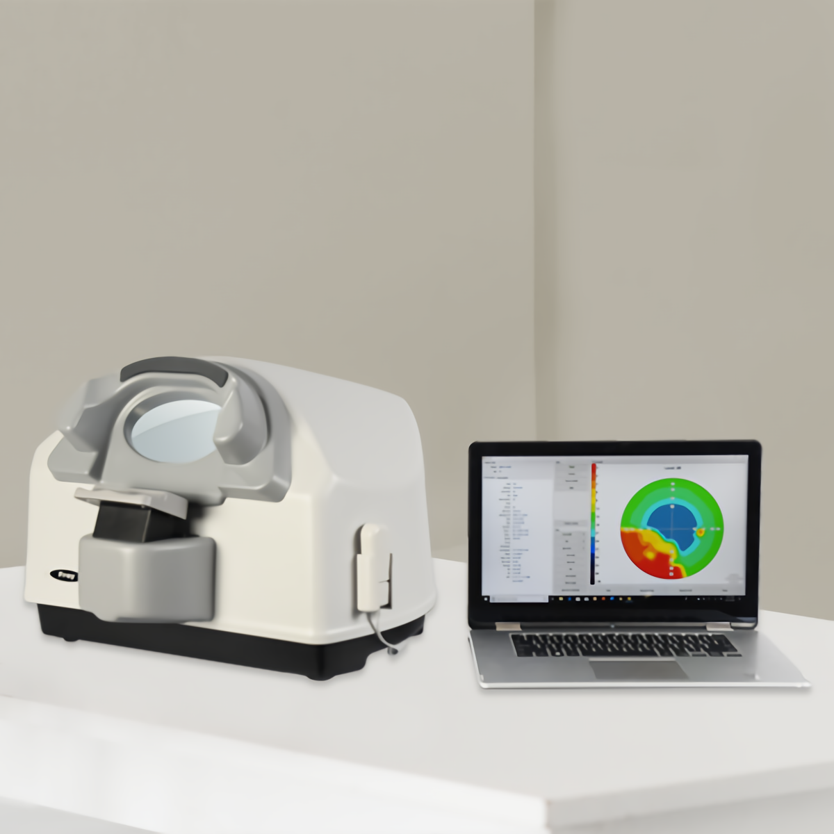
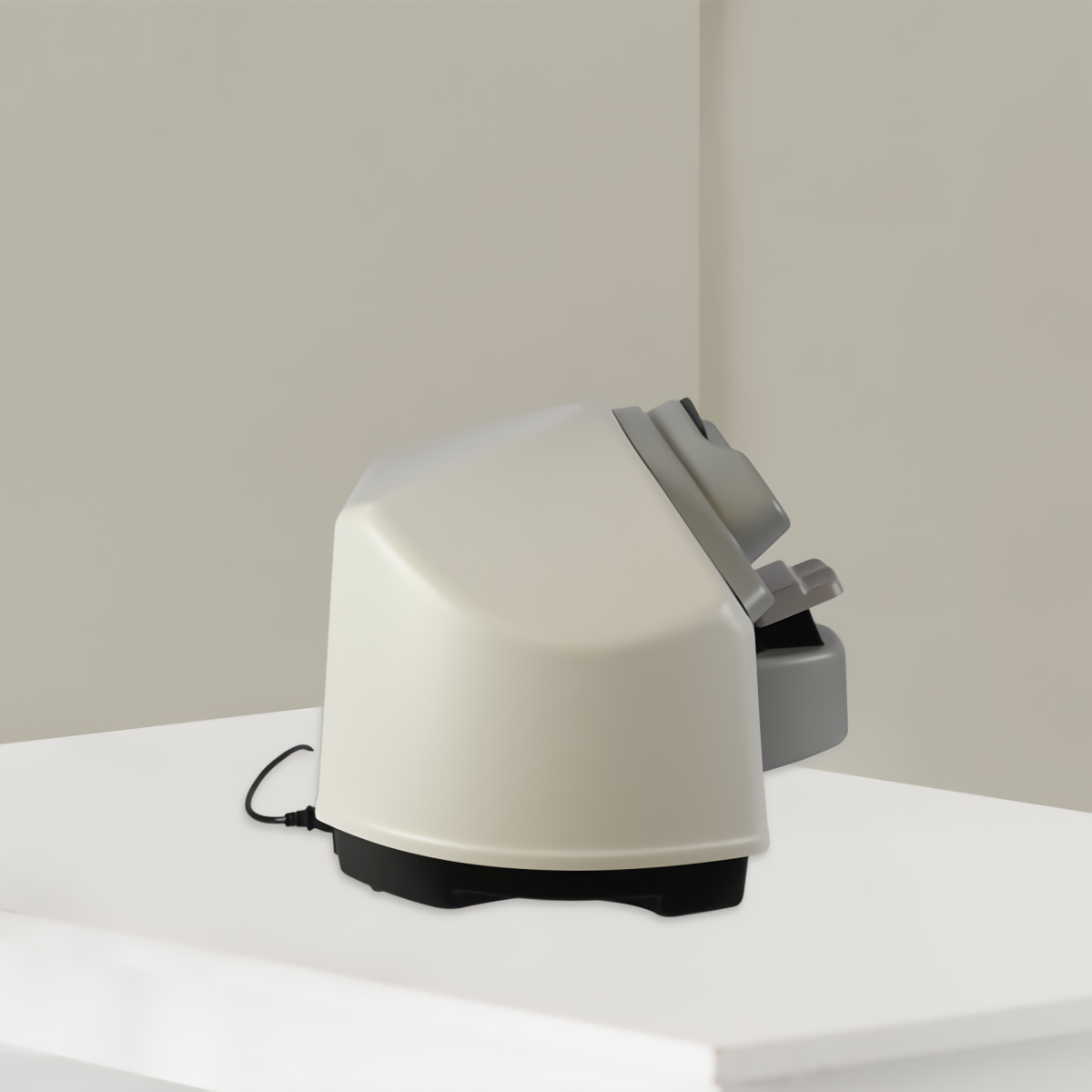
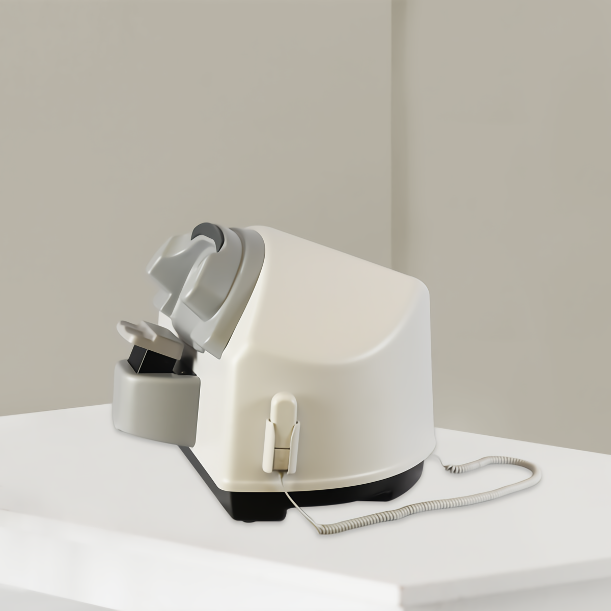
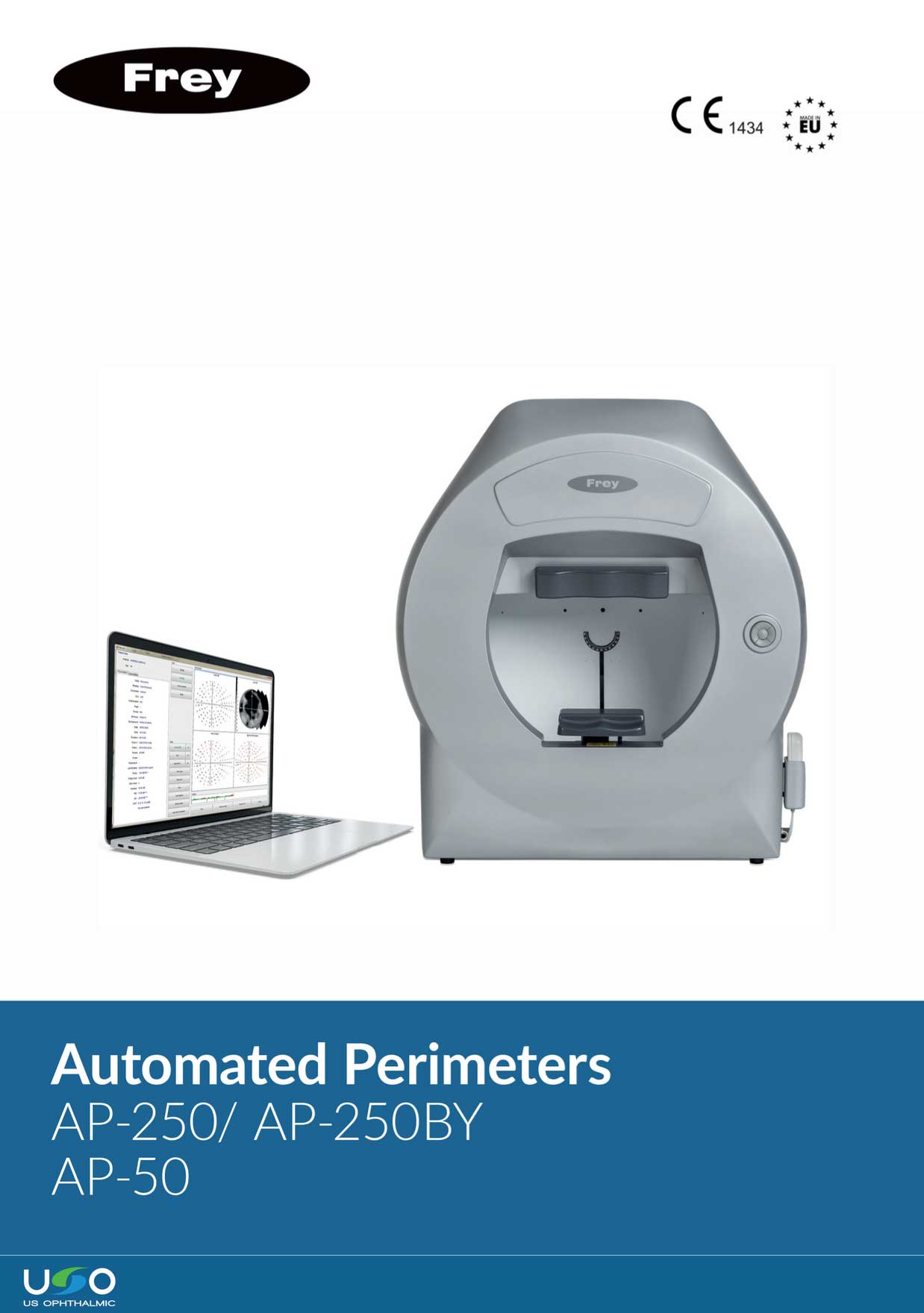
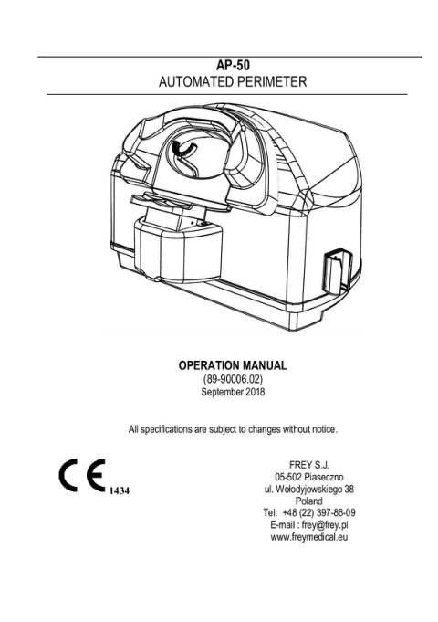
The AP-50 is a lightweight, fully equipped tabletop model — a modern automated static perimeter, ideal for glaucoma diagnosis and for the specific needs of occupational medicine and high-volume mobile clinics. The AP-50 uses rear LED projection of white-color stimuli and offers a wide range of strategies, test fields, and a complete set of parameters to ensure fast and accurate measurements. Fixation control is performed automatically via the integrated camera or through blind spot position monitoring. The built-in data analysis functions include regression analysis and standardized methods for presenting and printing examination results. The AP-50 perimeter can be used with any PC running the Windows operating system.

*PC not included
Talk to a specialist

Patient focus – greater comfort.

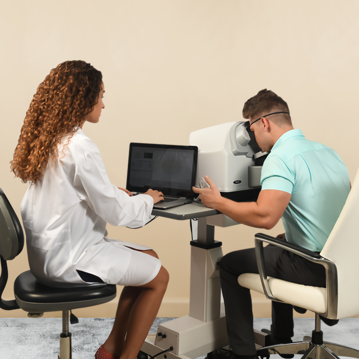
Visual field testing is a collaborative effort between the specialist and the patient. Perimetry is a team effort. A well-assisted patient, for whom comfort is maximized, improves the reliability of the visual field examination. This greater reliability translates into more precise and effective diagnosis and clinical management. In turn, it improves outcomes and satisfaction throughout the patient journey, which can extend over decades. Frey has provided greater patient comfort with a chin rest designed for comfort and stability. The patient’s head remains supported throughout the entire examination. The AP-50 features enhanced patient ventilation, which reduces stress and increases comfort.
Precise Results

The stimulating dome offers a high density of concentric dots. The enhanced stimulus control, combined with automated eye tracking, provides repeatable and precise examinations of the patient’s visual field loss.

Fast Test Times

Efficient test completion is ensured through fast detection and threshold strategies, as well as enhanced fixation methods. Patients with advanced visual field loss benefit from standard calibration support.

Operation Modes
Static Test Mode

The AP-50 emits stationary stimuli of variable luminance to determine the patient’s threshold. A faint light appears at a specific location and, if the patient does not perceive it, the stimulus intensity increases. Patient responses are compared to those of an individual from a control group of the same age. The test is defined by the test and strategy, white-on-white stimuli and Goldman III size. These are available for static testing, and the results are more reliable and of higher quality.

Binocular Testing

The AP-50 meets specific visual field testing requirements for drivers and occupational medicine. The measurement range covers up to 160 degrees in the temporal direction, and both eyes can be evaluated simultaneously.

Static Test Parameters

The background illumination color is white for all tests. Selecting the appropriate test strategy is essential, and we evaluate nearly twice as many points as our competitors for the same visual field. Additionally, the AP-50 offers an intelligent threshold that further enhances test accuracy.

Results Review – Static Perimeter

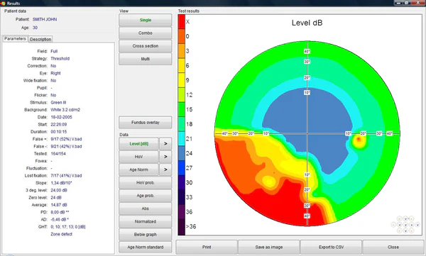
The “Results” screen is divided into groups of functional boxes. Detailed descriptions of these boxes are provided in the following section (Individual, Combined, Cross-sectional View, and Multiple).

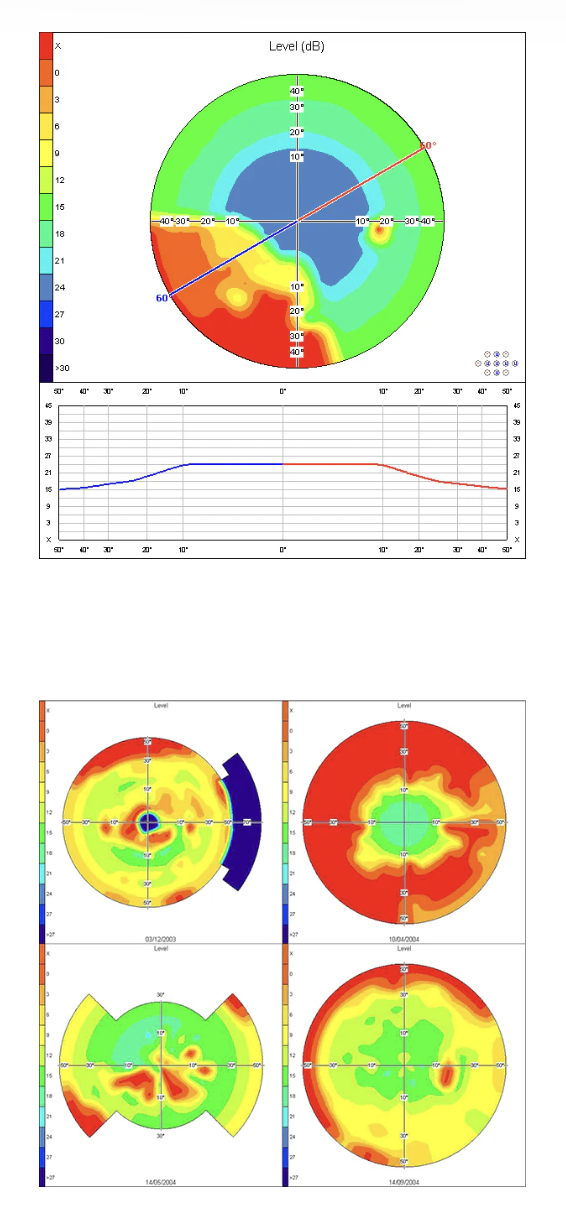
Cross-sectional view

A graphic map that divides the visual field into two parts, represented by the colors red and blue. This study facilitates the selection of the cut angle and allows sensitivity to be measured in decibels in all directions of the visual field up to 60˚.

Fundus Overlay Function

This is a unique approach that allows selective evaluation of retinal areas. Stimulus locations are established from the patient’s fundus image, while the area and density of test points are configured manually.

Fundus Overlay View

The Automated Perimeter software allows users to overlay examination results with fundus images captured by the fundus camera. To obtain the overlay, after selecting the “Fundus Overlay” option, load the fundus image from a file or select the image from those already saved. The next step is the selection of two reference points, which are used to calculate the positions of the examination results. These points are the fovea and the blind spot. Press the button labeled “Fovea” and click on the position of the fovea on the fundus image. Then, press the button labeled “Blind Spot” and click on the position of the blind spot on the fundus image. The examination results are now displayed over the fundus image. The overlay can subsequently be saved using the “Save Overlay” option.

Intuitive Software

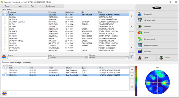
El AP-50 was designed with a focus on both the patient and the clinical specialist. The Frey perimeter software is intuitive and easy to use, enabling efficient and effective operation by different professionals. The interactive menus offer comprehensive information and efficient operation, reducing preparation, review, and printing time for examinations. This platform supports clinical excellence with increased patient throughput and greater efficacy.

Version information
Add new patient
Edit patient data
Start new examination
Results review
Comparison function
Regression analysis
Test editor
Export/import function

Dual main screen option

This feature offers two main screen options; it is streamlined for use in the testing room by the operator, allows customized test selection, constitutes a standardized testing procedure, and includes a complete interface for reception areas or medical offices.

Simple Menu Customization

The simplified menu is fully customizable, and all test parameters can be configured according to the customer’s needs. The clinical workflow is also optimized, allowing standardization of the testing procedure. Furthermore, the software is very easy to use, with minimal requirements.

Fixation Control

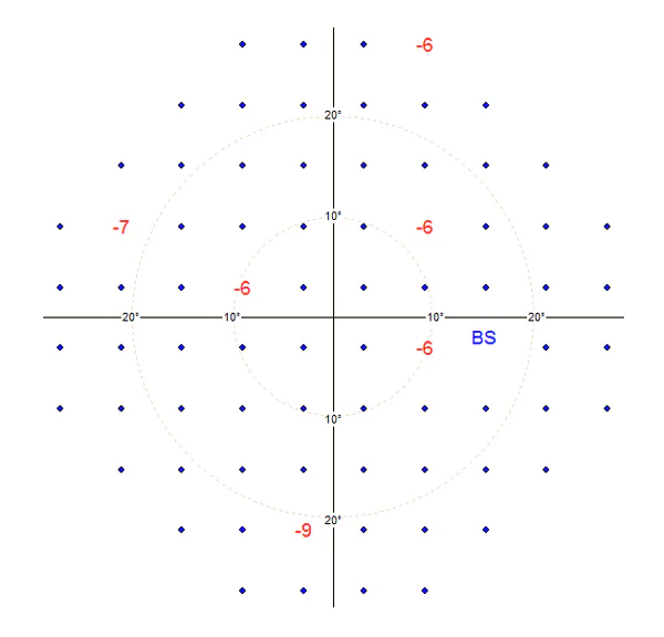
The Automated Perimeter AP-50 features two fixation control mechanisms. Heijl-Krakau Method A classic method that verifies the position of the blind spot by evaluating a total of 11 random points to ensure correct eye positioning. Ocular Position Method The AP-50 facilitates fixation analysis through a video camera that enables continuous monitoring of the pupil. The advantage of this method is that it rejects patient responses when fixation is not present.

Reliability

The automatic fixation control, with integrated camera and automatic eye tracking, assists both the patient and the specialist. This increases the repeatability and reliability of data capture.


Automatic pupil detection

When the automatic fixation monitoring method is used, the software detects the current position of the eye. It is not necessary to keep the eye centered on the image during the test, although it is essential that the patient always looks at the fixation point. Eye positioning control can be performed via the screen, the joystick, or the software. Additionally, the chin rest can be moved up and down.

Advanced Techniques

En el pasado, el glaucoma solo se diagnosticaba en etapas avanzadas. Actualmente, las nuevas tecnologías permiten la detección temprana de esta condición.

Flicker (Critical Flicker Fusion)

Glaucoma affects sensitivity to flickering lights.
The CFF analyzes the patient’s ability to perceive alternations of light and darkness across different areas of the visual field. It also measures early neuronal damage, detecting glaucoma in its initial stages.

Single Print

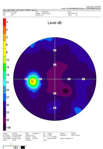
The AP-50 uses the simplest printing format, which contains a large graphic map representing the examination results. The print will be produced in the same format selected on the “Results” screen in “Single Mode”. When “Single Mode” is not selected, the dB level map in grayscale is printed by default. The advantage of the “Single Print” option is its high readability.

LED LIGHT SOURCE

* All AP-50 light sources are LED
* The system requires no maintenance
* Demonstrates high light source stability over time
* True white-on-white perimeter

Reliability

The reliable automatic fixation control, with integrated camera and automatic eye tracking, assists both the patient and the specialist, increasing repeatability and reliability in data capture.

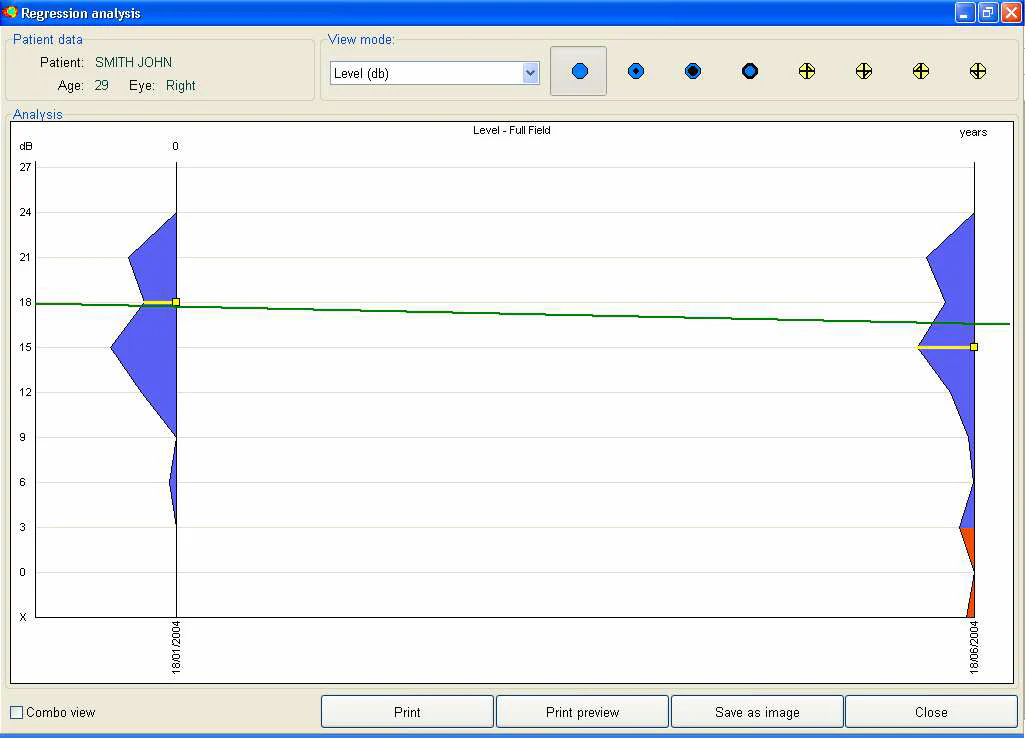
Regression analysis – static perimetry

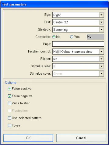
The AP software is equipped with options that allow observing changes in the patient’s vision in several ways. The first option is the simple comparison of results, described in the previous section. The second, more advanced option is the regression analysis, which helps the user observe the progression or regression of vision over time.
At least two examination results from the patient are required to perform the regression analysis. The examinations must be performed using the same strategy, but on different days.
The patient’s name, age, and examined eye are displayed in the upper left of the screen. A selection box in the upper center allows choosing one of five parameters to calculate and display the regression graph. The following can be selected:

Decibel level
Hill of vision – decibel level
Pattern Defect (PD)
Average Defect (AD)

The selection box is available when “Individual Mode” is selected (in the lower right corner of the screen). “Combined Mode” displays all five regression graph modes together.

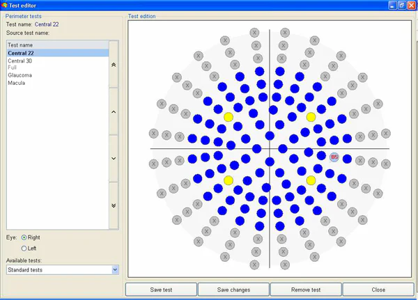
Test Editing

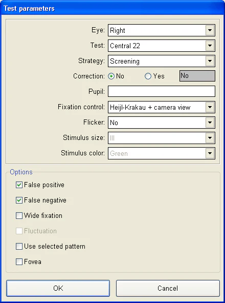
This function allows defining a custom test field. It is especially useful when only part of the patient’s visual field needs to be examined, for example, the area where field loss was observed in previous exams. Using a custom field can reduce patient discomfort and examination time.
Before starting the editing process, the affected eye (right or left) and the name of the test field to be used as default must be selected. Fields available by default:

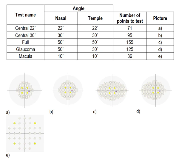
Central 22˚
Central 30˚
Full
Glaucoma
Scotoma

The selection box is available when “Single Mode” is selected (in the lower right corner of the screen). “Combined Mode” displays all five regression graph modes together.

Probability map of the hill of vision

The “probability map of the hill of vision” displays the likelihood of hill of vision defects at each point of the visual field. Probabilities are calculated based on the differences between the theoretical and the calculated hills. The lower the probability, the greater the visual field defect.

Complete Analysis Modes

La confiabilidad y funcionalidad del AP-50 se ven mejoradas por modos de análisis completos basados en estadísticas de población mundial.
Los mapas sombreados cuentan con una funcionalidad 3D mejorada.

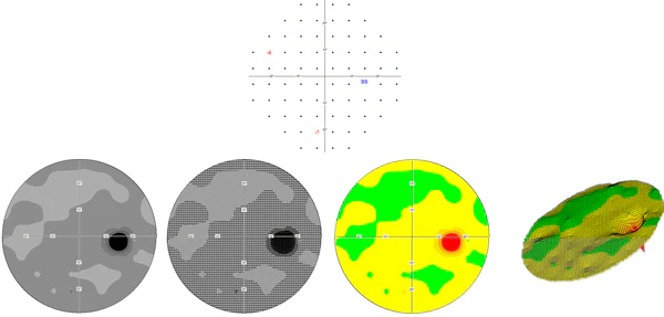
Age-Normal Map

The “Age-Normal Map” displays the differences between age-based normal values and the values obtained during the test. In this way, it reflects the difference between the theoretical dB level map and the measured value.
The “Age-Normal Map” can be displayed in the following formats: numeric, grayscale, color scale, pattern scale, and 3D graph.

Multiple Testing Capabilities

The automated perimetry technology from Frey offers complete flexibility for the specialist and the patient. It allows testing for glaucoma, full field, and flicker. This flexible platform addresses clinical challenges ranging from glaucoma to neurology and beyond.

Standard Tests – Static Perimetry

Standard tests are available by default in the AP software, and each one is described in the tables and images below.

Networks

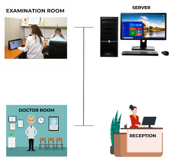
The AP-50 consistently delivers excellence in visual field measurement and analysis. This critical information can be integrated into the computer network; multiple perimeters can share a single database, supporting large clinics and high patient throughput. The automatic backup function ensures the security of patient and clinical data throughout the entire process. Frey takes pride in offering access to the highest quality technical support and service, anytime and anywhere.

The AP-50 software was designed to operate in a PC network environment.
It can be installed on multiple PCs at no additional licensing cost.
All PCs can share a single database.
A centralized server can be used for database storage.
An automatic backup function is available to ensure data security.

DICOM Export

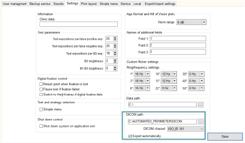
Each examination result can be stored in DICOM format directly from the data review screen. Additionally, automatic DICOM storage to any network location can be configured in the service settings.


Specifications

Manuals & Catalogs

Catalogs

Frey Perimeter AP-50

Catalogs

Frey Perimeter AP-50

Manuals

Frey Perimeter AP-50